1,144
Views
12
CrossRef citations to date
0
Altmetric
Review

The effect of mechanical strain on soft (cardiovascular) and hard (bone) tissues

Common pathways for different biological outcomes

, , , &
Pages 165-173 | Received 03 Oct 2012, Accepted 27 Nov 2012, Published online: 03 Jan 2013

Figures & data

Figure 1. Compact bone microanatomy (acid fuchsin-light green staining 1,200× oil).

Figure 1. Compact bone microanatomy (acid fuchsin-light green staining 1,200× oil).

Figure 2. Schematic representation of the main molecular pathways involved in bone responses to mechanical strain.
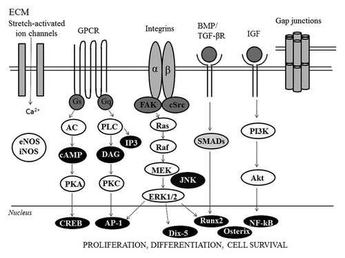
Figure 2. Schematic representation of the main molecular pathways involved in bone responses to mechanical strain.

Figure 3. Blood vessels microanatomy (hematoxilin-eosin staining 430×).

Figure 3. Blood vessels microanatomy (hematoxilin-eosin staining 430×).

Figure 4. Mechanical and hemodynamic forces associated with blood flow: Shear stress (τ) is parallel to the vessel wall and represents the frictional force that blood flow exerts mainly on the endothelial surface of the vessel wall. Instead, cyclic stretch (ρ) is the stress perpendicular to the vessel wall and represents the circumferential deformation of the blood vessel wall during distension and relaxation of the recurring cardiac cycle.
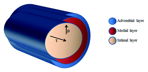
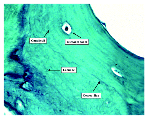
Figure 4. Mechanical and hemodynamic forces associated with blood flow: Shear stress (τ) is parallel to the vessel wall and represents the frictional force that blood flow exerts mainly on the endothelial surface of the vessel wall. Instead, cyclic stretch (ρ) is the stress perpendicular to the vessel wall and represents the circumferential deformation of the blood vessel wall during distension and relaxation of the recurring cardiac cycle.
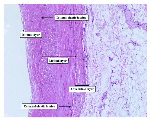
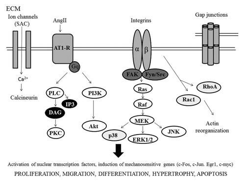
Figure 5. Main molecular pathways involved in mechanotransduction in vascular cells.

Figure 5. Main molecular pathways involved in mechanotransduction in vascular cells.

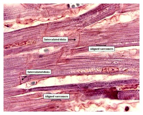
Figure 6. Heart microanatomy showing the intercalated disks (Phosphotungstic acid hematoxylin staining 1,200× oil).

Figure 6. Heart microanatomy showing the intercalated disks (Phosphotungstic acid hematoxylin staining 1,200× oil).

Reprints and Corporate Permissions

Please note: Selecting permissions does not provide access to the full text of the article, please see our help page How do I view content?

To request a reprint or corporate permissions for this article, please click on the relevant link below:

Academic Permissions

Please note: Selecting permissions does not provide access to the full text of the article, please see our help page How do I view content?

Obtain permissions instantly via Rightslink by clicking on the button below:

If you are unable to obtain permissions via Rightslink, please complete and submit this Permissions form. For more information, please visit our Permissions help page.