1,138
Views
15
CrossRef citations to date
0
Altmetric
Commentary

Protein tyrosine phosphatase PTP1B in cell adhesion and migration

, , &
Pages 418-423 | Received 05 Aug 2013, Accepted 04 Sep 2013, Published online: 12 Sep 2013

Figures & data

Figure 1. PTP1B localization and function on adhesion and motility in polarized, migrating cells. (A and B) GFP-PTP1B (green label) localizes at the ER and overlaps with vinculin adhesions (red label) at the cell periphery. (C and D) Microtubules (blue label) contribute to position the substrate trap GFP-PTP1BDA (green label) in paxillin adhesions (red label), where it binds to substrates and accumulates in small puncta (arrowheads in D). Arrows in (A and C) point the leading edge. (E) ER tubules randomly contact the plasma membrane outside adhesions in a microtubule (MT)-dependent manner. Membrane-associated, inactive Src (blue spheres, (p)Src), is targeted by ER-bound PTP1B at the pTyr-529, causing its dephosphorylation (light blue spheres, de(p)Src) and priming the kinase for activation at the plasma membrane. (F) MT-dependent positioning of ER-bound PTP1B over peripheral adhesions facilitates the activity of PTP1B on multiple substrates, including α-actinin, paxillin, and Src. (G) By targeting tyrosine 12 on α-actinin (in red) PTP1B contributes to enhance α-actinin binding to actin and to reinforce integrin/cytoskeleton linkages. By targeting Src regulatory pY529, PTP1B promotes Src activation and signaling through Rac1 and RhoA. During the protrusion phase, PTP1B promotes Rac1 activity and inhibits RhoA activity. Scale bars, 15 µm. Magnifications (B and D) are 400x of the original size.
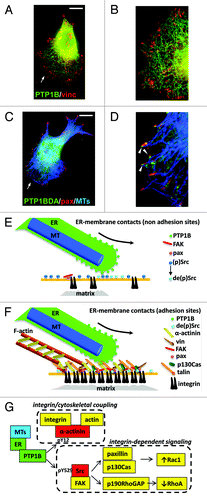
Figure 1. PTP1B localization and function on adhesion and motility in polarized, migrating cells. (A and B) GFP-PTP1B (green label) localizes at the ER and overlaps with vinculin adhesions (red label) at the cell periphery. (C and D) Microtubules (blue label) contribute to position the substrate trap GFP-PTP1BDA (green label) in paxillin adhesions (red label), where it binds to substrates and accumulates in small puncta (arrowheads in D). Arrows in (A and C) point the leading edge. (E) ER tubules randomly contact the plasma membrane outside adhesions in a microtubule (MT)-dependent manner. Membrane-associated, inactive Src (blue spheres, (p)Src), is targeted by ER-bound PTP1B at the pTyr-529, causing its dephosphorylation (light blue spheres, de(p)Src) and priming the kinase for activation at the plasma membrane. (F) MT-dependent positioning of ER-bound PTP1B over peripheral adhesions facilitates the activity of PTP1B on multiple substrates, including α-actinin, paxillin, and Src. (G) By targeting tyrosine 12 on α-actinin (in red) PTP1B contributes to enhance α-actinin binding to actin and to reinforce integrin/cytoskeleton linkages. By targeting Src regulatory pY529, PTP1B promotes Src activation and signaling through Rac1 and RhoA. During the protrusion phase, PTP1B promotes Rac1 activity and inhibits RhoA activity. Scale bars, 15 µm. Magnifications (B and D) are 400x of the original size.

Table 1. PTP1B substrates involved in cell adhesion and migration