Figures & data
Figure 1 Neural crest cell migration. Neural crest (NC) cells (blue) emerge from the dorsal neuroepithelium and migrate extensively throughout the embryo. The cephalic NC cells mainly migrate under the skin and toward the ventral portions of the face with some subpopulations migrating further ventrally toward the heart and along gut. The trunk NC cells mostly invade the ventral regions of the trunk and colonize the skin.
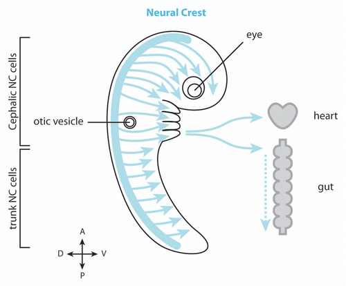
Figure 2 Migration of cephalic NC cells in Xenopus and chick embryos. (A) In Xenopus, NC cell migration start as a loose cell sheet (top part) and progressively turns into a cell streaming composed of mesenchymal cells (bottom part). (B) In chick, NC cells migrate as mesenchymal cells and form chains. In both models cells are polarized by interactions with other NC cells (red) and maintained as a dense group by the presence of inhibitors defining the borders of the NC routes (shades of orange). High cell density leads to directional movement while isolated cells exhibit poor directionality (sinuous path).
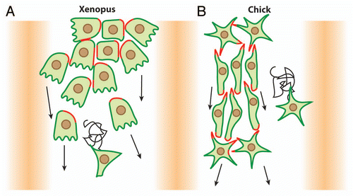
Figure 3 Xenopus cephalic NC cells are polarized by contact-inhibition of locomotion. (A) Contact-Inhibition of Locomotion is triggered by cell interactions. Cells are polarized according to their cell-cell contacts; free edge is in green, cell contacts are in red. (B) Cell-cell interactions are mediated by N-Cadherin and Wnt/PCP signaling. N-Cadherin is required for a local inhibition of Rac1 at the cell junctions and Wnt/PCP induces an increase of RhoA activity at the contact. Both N-Cadherin and Wnt/PCP maintain low Rac1 and high RhoA activities at the cell-cell contact which restrict Rac1 activity at the free edge.
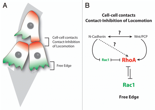
Figure 4 Neural crest cells exhibit solitary and collective behaviors. Comparison between epithelial cells, cephalic, enteric and trunk NC cells. For each cell population their epithelial or mesenchymal phenotype and the type of cell-cell contacts are indicated. Motility at the group level means that non-motile cells can be pulled by adjacent motile cells while motility at the single cell level indicates that each cell is properly motile. Response to external signals at the group level means that the ability to respond to these signals depends on the fact that cells are part of a collective as opposed to a situation where each cell responds to external cues independently. Such collective guidance has been shown for Xenopus and mouse cephalic NC cellsCitation13,Citation46 and is therefore likely to apply to other cephalic crests in other species. CNC, cephalic NC cells; TNC, trunk NC cells.
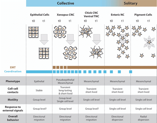
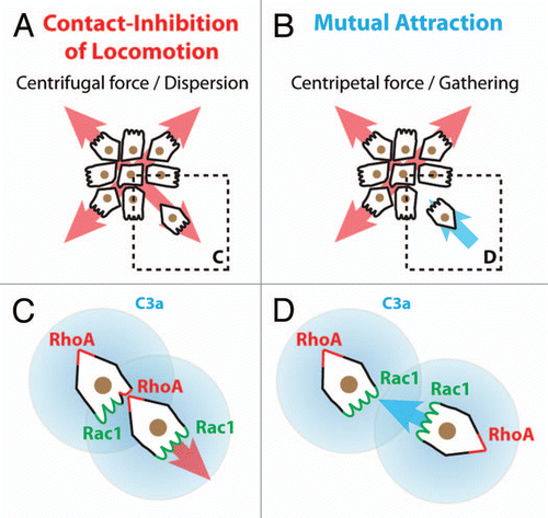
Figure 5 Collectiveness of Xenopus cephalic neural crest cells is maintained by mutual attraction based on autocrine C3a/C3aR signaling. (A) Contact-Inhibition of Locomotion (CIL) polarizes cells toward the cell-free space and therefore acts as a centrifugal force leading to dispersion. (B) Mutual attraction driven by chemoattractant C3a acts as centripetal force promoting gathering of cells. (C) Cell-cell interactions through CIL polarize the distribution and activity of small GTPases within the cells. (D) C3a/C3aR signaling promotes Rac1 activity in cells that have recently left the group. This leads to repolarization and gathering.