Figures & data
Figure 1. Analyses of SCF expression in T-ALL mice. To assess whether SCF expression was upregulated in the leukemic environment in our model, we performed ELISA to determine SCF concentration in PB serum and BM samples from leukemia and control mice 10 d after transplantation (A and B). The expression of SCF in BM, spleen and blood cells as well as CD45.1+ BM cells and GFP+ BM cells was analyzed by real-time PCR (C). The level of SCF protein in leukemia and normal cells was determined by western blot (D).
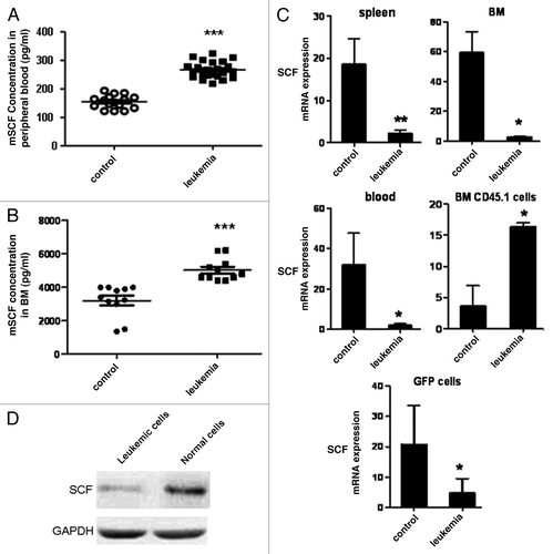
Figure 2. Gene profiling analyses of normal HSCs in leukemic environment. Normal HSCs were obtained from control and leukemia mice on day 10 after transplantation (A). The clustering map showed the genes differently expressed in normal HSCs between leukemia and control group. Red color meant the genes were upregulated while green color meant the genes were downregulated (B). Schematic representation of the analyses strategies integrating expression profiles obtained from two independent experiments (C).
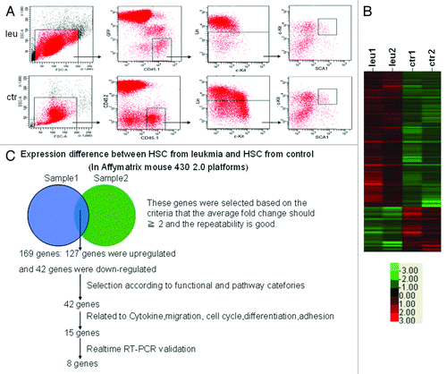
Figure 3. Validation of the results of microarray by real-time PCR. Real-time RT-PCR was used to validate the expression pattern of eight genes using specific murine primers detailed in (A). Quantitative analysis of Hes1 and p21 expression in normal HSCs and HPCs in leukemia and control mice was shown in (B and C). Data are shown as means ± SD *, p < 0.05, statistical analysis was performed using Student’s t-test.
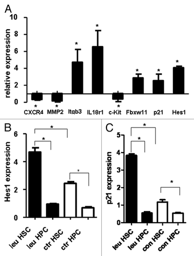
Figure 4. In vitro CFC study on Hes1-transduced HSPCs and HSCs. Infection rates were assessed using transduction of 293T cells. After infection, the cells were analyzed with fluorescence microscope (A) and GFP+ cells were sorted for the expression of Hes1 and other related genes (B). Lin- cells (C) and LSK cells (D) of B6.SJL mice were transduced with Hes1 or control retrovector. Successfully transduced (GFP+) cells were enriched by FACS sorting and plated in standard methylcellulose-containing CFC assays. Data are shown as means ± SD ***, p < 0.0001, statistical analysis was performed using Student’s t-test.
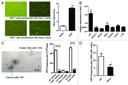
Figure 6. HSCs/HPCs with Hes1 overexpression were preserved under leukemic environment. The Hes1-transduced mouse model is described in detail in Materials and Methods. On day 10 after transplantation, the absolute number and proportion of normal CD45.1+GFP+Lin- cells (A), CD45.1+ GFP+ HSPCs (B), CD45.1+ GFP+ HSCs (C) were analyzed with BD FACS Aria II.
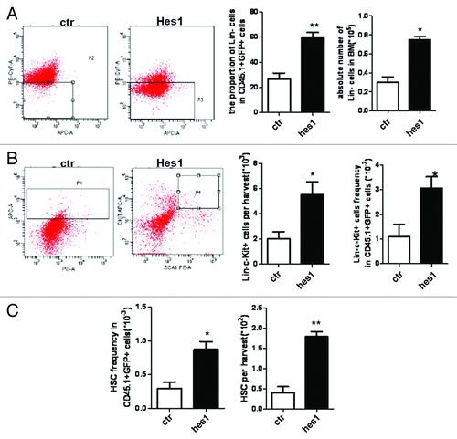
Figure 7. The function and cell cycle analyses of HSPCs in Hes1 and control model. On day 10 after transplantation, GFP+CD45.1+ cells were sorted for colony-forming cell (CFC) assay (A). The cell cycle of CD45.1+ GFP+ HSPCs was analyzed with Hoechst/PY (B).
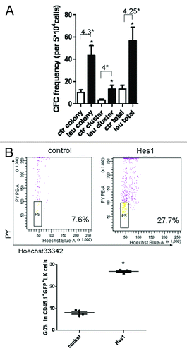
Table 1. The sequences of primers
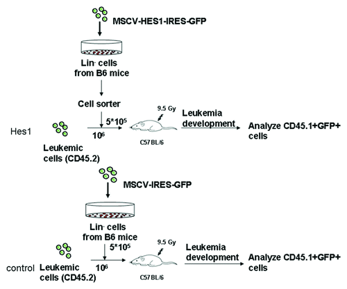
Figure 5. Establishment of Hes1-transduced mouse model. Lin- cells from B6.SJL mice at the age of 6–8 wk were transduced with either MSCV-Hes1-IRES-GFP or blank plasmid for 72 h. After transduction, Hes1-GFP+ or blank-GFP+ cells (CD45.1) were sorted and then injected into lethally irradiated C57BL/6J (CD45.2) by tail vein at a quantity of 2 × 10⁵ cells per mouse with 106 cells of Notch1-induced leukemia cells (CD45.2).