2,638
Views
85
CrossRef citations to date
0
Altmetric
Article Addendum

Tunneling Nanotubes

A new paradigm for studying intercellular communication and therapeutics in cancer

, , , , , & show all
Pages 399-403 | Published online: 01 Jul 2012

Figures & data

Figure 1. Tunneling nanotubes (TnTs) connecting mesothelioma cells (MSTO-211H cell line). All images were taken with Leica TCS SP5 with 40x/1.25NA or 100x/1.4NA oil objectives. (A) TnT connecting MSTO-211H cells, contrasted with shorter, adherent lamellopodia and actin stress fibers in the background. Blue = DAPI, green = actin (phalloidin stain), red = mitochondria (MitoTracker Red). (B) TnTs connecting MSTO-211H cells. Blue = DAPI, green = Golgi apparatus (GM130 antibody), red = mitochondria (MitoTracker Red). (C) TnT between MSTO-211H cells. Blue = Hoechst 3342, red = MitoTracker Red. (D) Ultrafine TnT connecting MSTO-211H cells. Blue = DAPI, red = RFP.
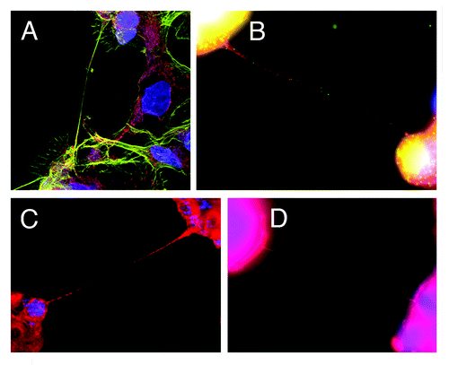
Figure 1. Tunneling nanotubes (TnTs) connecting mesothelioma cells (MSTO-211H cell line). All images were taken with Leica TCS SP5 with 40x/1.25NA or 100x/1.4NA oil objectives. (A) TnT connecting MSTO-211H cells, contrasted with shorter, adherent lamellopodia and actin stress fibers in the background. Blue = DAPI, green = actin (phalloidin stain), red = mitochondria (MitoTracker Red). (B) TnTs connecting MSTO-211H cells. Blue = DAPI, green = Golgi apparatus (GM130 antibody), red = mitochondria (MitoTracker Red). (C) TnT between MSTO-211H cells. Blue = Hoechst 3342, red = MitoTracker Red. (D) Ultrafine TnT connecting MSTO-211H cells. Blue = DAPI, red = RFP.

Figure 2. Tunneling nanotubes serve as a conduit for intercellular transport of organelles, include mitochondria. Numerous mitochondria were visualized in transit between MSTO-211H (biphasic mesothelioma) cells stained with MitoTracker Red. Images were captured using Leica inverted microscope, 100x/1.4NA oil objective.
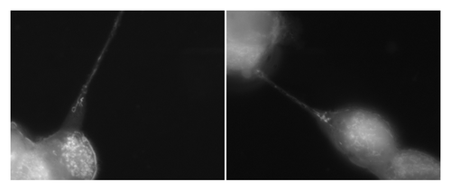
Figure 2. Tunneling nanotubes serve as a conduit for intercellular transport of organelles, include mitochondria. Numerous mitochondria were visualized in transit between MSTO-211H (biphasic mesothelioma) cells stained with MitoTracker Red. Images were captured using Leica inverted microscope, 100x/1.4NA oil objective.