Figures & data
Figure 1. Histologically confirmed hypertrophic actinic keratoses on right facial side of an elderly woman on a background of dermatoheliosis.
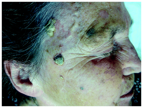
Figure 2. Field cancerization on scalp of an elderly man, with multiple actinic keratoses and histologically confirmed SCCs.
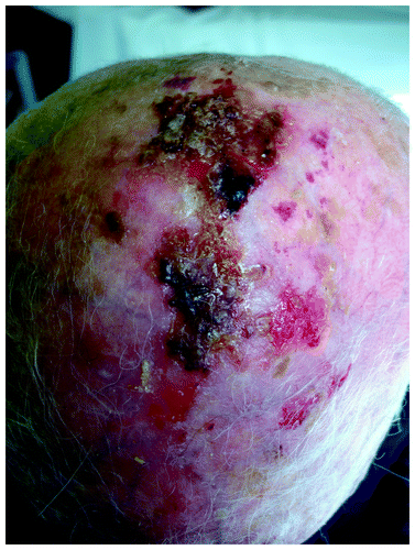
Figure 3. Histologically confirmed Bowen’s disease at right dorsal hand of an elderly patient existing for many years with a recently growing histologically confirmed Bowen’s carcinoma at the base of the thumb.
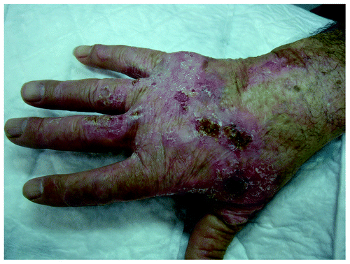
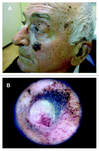
Figure 4. (A) Histologically confirmed nodular BCC in left preauricular area of a male patient. (B) Dermoscopic picture on same patient, showing central necrosis, maple leaf-like structures, blue-ovoid nests, blue-gray globules, and arborising vessels.
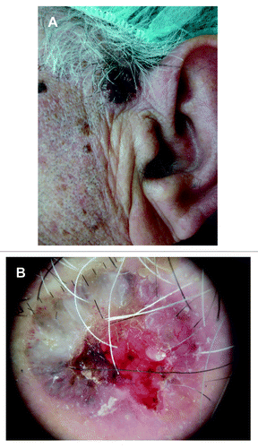
Figure 5. Histologically confirmed SCC on right helix of an elderly man.
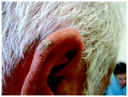
Figure 6. Histologically confirmed SCC on left temporal area of an elderly man, multiple actinic keratoses on left ear and lentigo maligna on his left cheek.
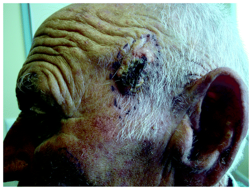
Figure 7. Multiple lentigines senilis on face of an elderly woman and a seborrheic keratosis on her left cheek.
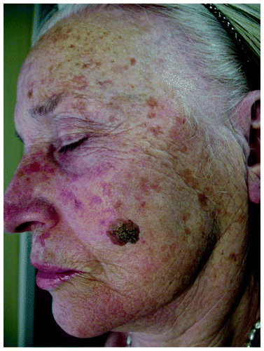
Figure 8. (A) Histologically confirmed lentigo maligna melanoma on left cheek of an elderly male patient, arising from a lentigo maligna after many years. (B) Dermoscopic picture of LMM on same patient.