Figures & data
Figure 1 Pilosebaceous unit in facial skin of acne patients. Faintly hypertrophic sebaceous gland are observed. Dilated capillaries and perivascular lymphocytes (A and C) are early signs of inflammatory process in acne-involved skin. Dilated plugged orifice of hair follicle—sign of acne comedo (B).
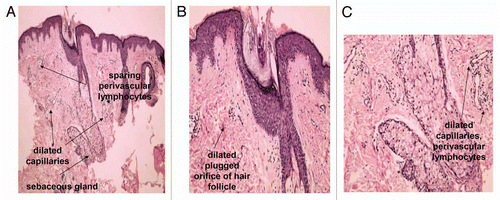
Figure 2 Regulation of the biological function of human sebaceous gland cells. Schematic overview. [LXR: liver X receptors, PPAR: peroxisome-proliferator activated receptors].
![Figure 2 Regulation of the biological function of human sebaceous gland cells. Schematic overview. [LXR: liver X receptors, PPAR: peroxisome-proliferator activated receptors].](/cms/asset/8f9286f3-bd7d-4a9c-8b03-ad783d46aa4d/kder_a_10913900_f0002.gif)
Figure 3 Localisation of CRH immunostaining in the sebaceous gland of acne patients (A) and in the normal skin of healthy controls (B). Very intensive expression of the gene in all types of sebocytes—basal, differentiating and mature cells—and keratinocytes of ductus seboglandularis in acne skin is shown (A) (x400); significant weaker and dependent upon sebocytes differentiation stage immunoreaction of sebaceous gland in normal skin (B) (x400).
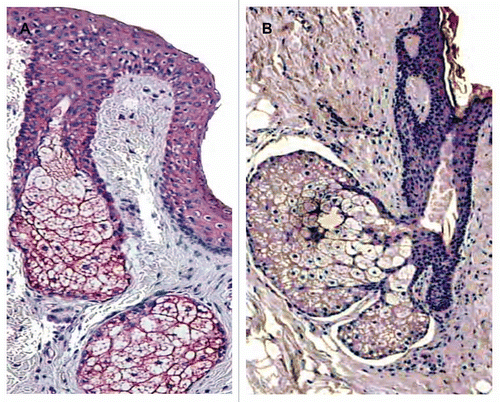
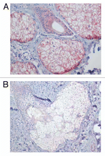
Figure 4 Localisation of COX-2 immunostaining in the sebaceous gland of acne patients (A) and in the normal skin of healthy controls (B). Very strong immunoreaction of the gene within the sebaceous gland of acne skin, especially, in undifferentiated and early differentiated sebocytes is seen (A) (x400). Weak immunoexpression of COX-2 in the sebaceous gland and ductal cells of healthy skin (B) (x400).