1,981
Views
71
CrossRef citations to date
0
Altmetric
Research Paper

A genome-wide approach reveals novel imprinted genes expressed in the human placenta

, , , , , , , , , , , , , , & show all
Pages 1079-1090 | Published online: 16 Aug 2012

Figures & data

Table 1. Summary of newly identified imprinted genes

Figure 1. Sequencing results of SNPs in the human ZFAT and ZFAT-AS1 genes. Imprinted expression was deduced by comparing genotypes of gDNA, cDNA and maternal gDNA, in placenta using SNPs rs rs3739423 (A), rs894344 (B) and rs894343 (C) and in lymphocytes (D).
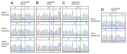
Figure 1. Sequencing results of SNPs in the human ZFAT and ZFAT-AS1 genes. Imprinted expression was deduced by comparing genotypes of gDNA, cDNA and maternal gDNA, in placenta using SNPs rs rs3739423 (A), rs894344 (B) and rs894343 (C) and in lymphocytes (D).

Figure 2. Sequencing results of SNPs in the murine (A) and bovine (B) Zfat genes. Imprinted expression was deduced by comparing genotypes of gDNA, cDNA and maternal gDNA, in placenta and other tissues.
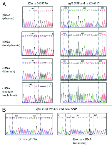
Figure 2. Sequencing results of SNPs in the murine (A) and bovine (B) Zfat genes. Imprinted expression was deduced by comparing genotypes of gDNA, cDNA and maternal gDNA, in placenta and other tissues.
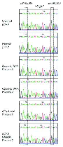
Figure 3. Sequencing results of SNPs in the murine Magi2 gene. Imprinted expression was deduced by comparing genotypes of gDNA, cDNA and maternal gDNA, in placenta and other tissues.

Figure 3. Sequencing results of SNPs in the murine Magi2 gene. Imprinted expression was deduced by comparing genotypes of gDNA, cDNA and maternal gDNA, in placenta and other tissues.

Figure 4. Real-time RT-PCR of ZFAT in human placentas. Expression of the ZFAT gene was compared between Controls (n = 17), IUGR (pregnancies with intrauterine growth restriction) (n = 16), PE (pregnancies complicated with preeclampsia) (n = 15) and PE + IUGR (n = 5), after normalization by the SDHA housekeeping gene. *p = 0,02, ** p = 0,002 in a group vs. the control group.
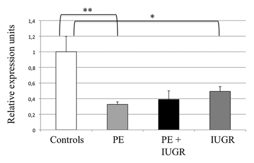
Figure 4. Real-time RT-PCR of ZFAT in human placentas. Expression of the ZFAT gene was compared between Controls (n = 17), IUGR (pregnancies with intrauterine growth restriction) (n = 16), PE (pregnancies complicated with preeclampsia) (n = 15) and PE + IUGR (n = 5), after normalization by the SDHA housekeeping gene. *p = 0,02, ** p = 0,002 in a group vs. the control group.

Figure 5. Immunohistochemistry to detect ZFAT on sections of human placentas. (A and B) (19 and 32 weeks of amenorrhea respectively) and (C) (fetal lung). (+) shows the detection with the primary antibody sc-87510 and (-) the negative control of detection without this antibody. Plain arrows show the strong labeling of endothelial cells while dotted arrows point to the syncytiotrophoblast labeling. (D) A western blot of human and mouse tissues revealed by an anti-ZFAT antibody. PBL stands for peripheral blood leukocytes whereas JEG-3 is a human choriocarcinoma cell line commonly used as a model of trophoblast.
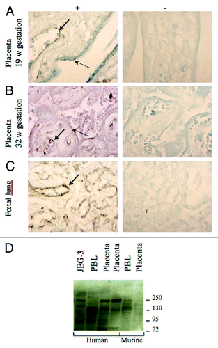
Figure 5. Immunohistochemistry to detect ZFAT on sections of human placentas. (A and B) (19 and 32 weeks of amenorrhea respectively) and (C) (fetal lung). (+) shows the detection with the primary antibody sc-87510 and (-) the negative control of detection without this antibody. Plain arrows show the strong labeling of endothelial cells while dotted arrows point to the syncytiotrophoblast labeling. (D) A western blot of human and mouse tissues revealed by an anti-ZFAT antibody. PBL stands for peripheral blood leukocytes whereas JEG-3 is a human choriocarcinoma cell line commonly used as a model of trophoblast.

Figure 6. Cloning and sequencing of PCR fragments obtained from bisulfite-treated placental gDNA in the promoter CpG island of the LIN28B gene. Black and white circles represent methylated and unmethylated cytosines respectively over the 32 CpG dinucleotides under study. SNPs are present within this fragment and allow the study of the allelic segregation and parental transmission.
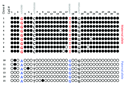
Figure 6. Cloning and sequencing of PCR fragments obtained from bisulfite-treated placental gDNA in the promoter CpG island of the LIN28B gene. Black and white circles represent methylated and unmethylated cytosines respectively over the 32 CpG dinucleotides under study. SNPs are present within this fragment and allow the study of the allelic segregation and parental transmission.
Supplemental material

Additional material

Download Zip (338.4 KB)