4,014
Views
58
CrossRef citations to date
0
Altmetric
Review

Recent advances in oral vaccine development

Yeast-derived β-glucan particles

, &
Pages 1309-1318 | Received 30 Jan 2014, Accepted 10 Feb 2014, Published online: 19 Feb 2014

Figures & data

Figure 1. Transmission electron micrograph (A) and scanning electron microscopy (B) images of ovalbumin-loaded β-glucan particles (De Smet et al.Citation60).
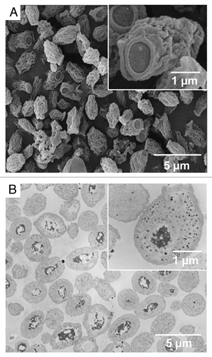
Figure 1. Transmission electron micrograph (A) and scanning electron microscopy (B) images of ovalbumin-loaded β-glucan particles (De Smet et al.Citation60).

Figure 2. Structure of yeast β-glucan (adapted from Volman et al.Citation112). Polymer of β-(1–3)-D-glycopyranosyl units with branching at β-(1–6)-D-glycopyranosyl units. S. cerevisiae structure consists of β-1–3 and small numbers of β-1–6 branches and β-1–6 linkages.
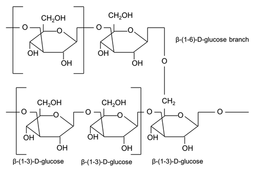
Figure 2. Structure of yeast β-glucan (adapted from Volman et al.Citation112). Polymer of β-(1–3)-D-glycopyranosyl units with branching at β-(1–6)-D-glycopyranosyl units. S. cerevisiae structure consists of β-1–3 and small numbers of β-1–6 branches and β-1–6 linkages.

Figure 3. Schematic representation of the pathways of β-glucan microparticle uptake. Beta-glucan microparticulate antigen uptake in mucosal inductive sites may occur through 3 different pathways: M-cells, intestinal epithelial cells, and dendritic cells. (Route 1) Non-migratory gut-resident CX3CR1-expressing dendritic cells protrude dendrites through epithelial tight junctions into the lumen to directly sample luminal antigen. (Route 2) Transcellular uptake of β-glucan microparticles across enterocytes by endocytosis. (Route 3) M-cells are specialized in antigen uptake because of their sparse glycocalyx, limited microvillus border, reduced enzymatic activity, and active transcytotic pathway. Transcytosis is a particular process by which M-cells endocytose antigens at their apical membrane, resulting in antigen transport into endosomal tubules, vesicles and large multivesicular bodies and finally exocytosis into the basolateral pocket. FAE, follicle-associated epithelium; SED, subepithelial dome; IFR, interfollicular region; HEV, high endothelial venule.
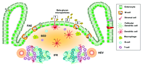
Figure 3. Schematic representation of the pathways of β-glucan microparticle uptake. Beta-glucan microparticulate antigen uptake in mucosal inductive sites may occur through 3 different pathways: M-cells, intestinal epithelial cells, and dendritic cells. (Route 1) Non-migratory gut-resident CX3CR1-expressing dendritic cells protrude dendrites through epithelial tight junctions into the lumen to directly sample luminal antigen. (Route 2) Transcellular uptake of β-glucan microparticles across enterocytes by endocytosis. (Route 3) M-cells are specialized in antigen uptake because of their sparse glycocalyx, limited microvillus border, reduced enzymatic activity, and active transcytotic pathway. Transcytosis is a particular process by which M-cells endocytose antigens at their apical membrane, resulting in antigen transport into endosomal tubules, vesicles and large multivesicular bodies and finally exocytosis into the basolateral pocket. FAE, follicle-associated epithelium; SED, subepithelial dome; IFR, interfollicular region; HEV, high endothelial venule.

Figure 4. (A) Activated dendritic cells may migrate to the mesenteric lymph nodes and process the β-glucan particles into peptides that are presented to T-cells. Presentation of β-glucan microparticulate antigen by dendritic cells via MHC class II molecules to CD4+ helper T-cells, leads to the activation, proliferation, and differentiation of antigen-specific Th17 and Th1 cells. Furthermore, dendritic cells and the intestinal epithelium produces cytokines such as BAFF, APRIL, and TGF-β1 that trigger the process of isotype switching and differentiation of IgA-committed B cells to IgA-producing plasma cells, which produce dimeric IgA. (B) Mature APCs and antigen-primed lymphoid cells will travel along the mesenteric lymph nodes, through the thoracic duct into the blood stream and end up at mucosal effector sites such as epithelia and lamina propria, where a cellular (effector T-lymphocytes) and humoral (secretory IgA-production by plasma cells) immune response is generated. CCR9 or CCR10 is expressed on gut-homing B-cells and T-cells which interact with CCL25 or CCL28 on the epithelium of the small or large intestine, respectively. IgA, immunoglobulin A; SC, secretory component; S-IgA, secretory IgA.
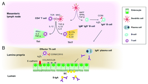
Figure 4. (A) Activated dendritic cells may migrate to the mesenteric lymph nodes and process the β-glucan particles into peptides that are presented to T-cells. Presentation of β-glucan microparticulate antigen by dendritic cells via MHC class II molecules to CD4+ helper T-cells, leads to the activation, proliferation, and differentiation of antigen-specific Th17 and Th1 cells. Furthermore, dendritic cells and the intestinal epithelium produces cytokines such as BAFF, APRIL, and TGF-β1 that trigger the process of isotype switching and differentiation of IgA-committed B cells to IgA-producing plasma cells, which produce dimeric IgA. (B) Mature APCs and antigen-primed lymphoid cells will travel along the mesenteric lymph nodes, through the thoracic duct into the blood stream and end up at mucosal effector sites such as epithelia and lamina propria, where a cellular (effector T-lymphocytes) and humoral (secretory IgA-production by plasma cells) immune response is generated. CCR9 or CCR10 is expressed on gut-homing B-cells and T-cells which interact with CCL25 or CCL28 on the epithelium of the small or large intestine, respectively. IgA, immunoglobulin A; SC, secretory component; S-IgA, secretory IgA.

Reprints and Corporate Permissions

Please note: Selecting permissions does not provide access to the full text of the article, please see our help page How do I view content?

To request a reprint or corporate permissions for this article, please click on the relevant link below:

Academic Permissions

Please note: Selecting permissions does not provide access to the full text of the article, please see our help page How do I view content?

Obtain permissions instantly via Rightslink by clicking on the button below:

If you are unable to obtain permissions via Rightslink, please complete and submit this Permissions form. For more information, please visit our Permissions help page.