Figures & data
Figure 1. Fiberd confocal fluorescence microscopy (FCFM) associated to a continuous aspiration system to assess the subpleural microcirculation in spontaneously breathing animals. The FCFM probe is sealed into a cylindrical chamber that is connected to an aspiration device (vacuum pump) providing a continuous depression of 25 cm H2O into the chamber. The extremity of the chamber, where the probe-tip emerges, is sealed to the lateral thoracic wall. The chest had been surgically prepared by removing the thoracic muscles layers up the internal intercostal muscle. The probe is then applied through the last muscle onto the pleural surface.
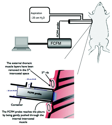
Figure 2. The subpleural capillary network observed with fiberd confocal fluorescence microscopy (FCFM) after IV injection of fluorescein. (A) The subpleural capillary network is easily recognized, and extra-alveolar vessels can be observed (*). (B) The intercapillary distance was measured on selected frames displaying typical capillary network. The ten maximal intercapillary distances were selected for the quantitative analysis, and are displayed as median ± interquartile range for each animal. Inter-individual variability, assessed by the inter-quartile range of the medians was [30–55] µm in the control group, and [42–66] µm in the elastase group. (C) The intercapillary distance was higher in the elastase-treated animals (median = 49.5µm [IQR (40.8 - 62.4)]) than in controls (median = 41.8µm [IQR (32.7 – 51.9)]) (p < 0.001, Mann-Whitney test).
![Figure 2. The subpleural capillary network observed with fiberd confocal fluorescence microscopy (FCFM) after IV injection of fluorescein. (A) The subpleural capillary network is easily recognized, and extra-alveolar vessels can be observed (*). (B) The intercapillary distance was measured on selected frames displaying typical capillary network. The ten maximal intercapillary distances were selected for the quantitative analysis, and are displayed as median ± interquartile range for each animal. Inter-individual variability, assessed by the inter-quartile range of the medians was [30–55] µm in the control group, and [42–66] µm in the elastase group. (C) The intercapillary distance was higher in the elastase-treated animals (median = 49.5µm [IQR (40.8 - 62.4)]) than in controls (median = 41.8µm [IQR (32.7 – 51.9)]) (p < 0.001, Mann-Whitney test).](/cms/asset/9415d21f-2e8b-453c-b4c8-14878bceac0a/kinv_a_10923471_f0002.gif)
Figure 3. The subpleural alveolar facets could be observed with fiberd confocal fluorescence microscopy (FCFM) after a few minutes of examination. (A) The ten maximal diameters were selected for the quantitative analysis. (B) The 10 measures per animal are displayed as median ± interquartile range. Inter-individual variability, assessed by the inter-quartile range of the medians was [84–106] µm in the control group, and [104–152] µm in the elastase group. (C) The alveolar facets diameters were significantly higher in the elastase-treated animals (median = 118.5µm [IQR (97.2 - 145.9)]) than in controls (median = 95.1µm [IQR (81.6 - 106.8)]) (p < 0.0001, Mann-Whitney test).
![Figure 3. The subpleural alveolar facets could be observed with fiberd confocal fluorescence microscopy (FCFM) after a few minutes of examination. (A) The ten maximal diameters were selected for the quantitative analysis. (B) The 10 measures per animal are displayed as median ± interquartile range. Inter-individual variability, assessed by the inter-quartile range of the medians was [84–106] µm in the control group, and [104–152] µm in the elastase group. (C) The alveolar facets diameters were significantly higher in the elastase-treated animals (median = 118.5µm [IQR (97.2 - 145.9)]) than in controls (median = 95.1µm [IQR (81.6 - 106.8)]) (p < 0.0001, Mann-Whitney test).](/cms/asset/b138351e-71aa-4f7b-aeaa-44177e8ade2b/kinv_a_10923471_f0003.gif)
Figure 4. The fluorescence intensity (FI) was measured on ten frames representative of the capillary network, excluding frames displaying large extra-alveolar microvessels. (A) The 10 measures per animal are displayed as median ± interquartile range. Inter-individual variability, assessed by the inter-quartile range of the medians was [789–1120] arbitrary units (A.U.) in the control group, and [424–951] A.U. in the elastase group. (B) The mean FI was significantly lower in the elastase-treated animals (mean ± SD = 764 ± 341 A.U.) than in controls (mean ± SD = 926 ± 258 A.U.) (p = 0,004, unpaired t-test)
![Figure 4. The fluorescence intensity (FI) was measured on ten frames representative of the capillary network, excluding frames displaying large extra-alveolar microvessels. (A) The 10 measures per animal are displayed as median ± interquartile range. Inter-individual variability, assessed by the inter-quartile range of the medians was [789–1120] arbitrary units (A.U.) in the control group, and [424–951] A.U. in the elastase group. (B) The mean FI was significantly lower in the elastase-treated animals (mean ± SD = 764 ± 341 A.U.) than in controls (mean ± SD = 926 ± 258 A.U.) (p = 0,004, unpaired t-test)](/cms/asset/ac717528-daf9-4f8a-b7eb-77b75e17bd9c/kinv_a_10923471_f0004.gif)
Figure 5. The mean interwall distance (MIWD) was used to assess pulmonary emphysema ex vivo. It was significantly higher in the elastase group (B) (median = 44.8 [IQR (43.7 – 45.3)]) than in controls (A) (median = 38.3µm [IQR (36.9 – 40.4)]) (p = 0.001, Mann-Whitney test).
![Figure 5. The mean interwall distance (MIWD) was used to assess pulmonary emphysema ex vivo. It was significantly higher in the elastase group (B) (median = 44.8 [IQR (43.7 – 45.3)]) than in controls (A) (median = 38.3µm [IQR (36.9 – 40.4)]) (p = 0.001, Mann-Whitney test).](/cms/asset/3c874dbd-b1d0-431b-8994-439c42df8cc1/kinv_a_10923471_f0005.gif)
Figure 6. The size of the subpleural alveolar facets diameters was correlated with the mean interwall distance (rs = 0.65 ; p = 0.016, Spearman correlation test).
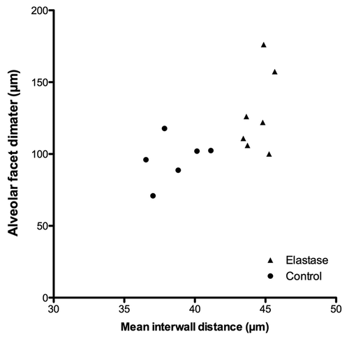
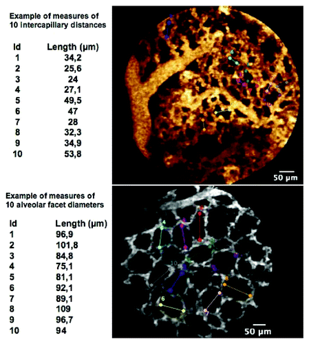
Figure 7. Examples of measurements of (top) intercapillary distances, and (bottom) alveolar facets diameters.