Figures & data
Figure 1. Intravital microscopy of tumors through imaging windows. (A) A 3D reconstruction of a murine orthotopic glioma (GL261-H2B Dendra2). The time-series show single cells migrating along the vessels. Nuclear localization of the photoprotein allows accurate single cell tracking. Green: tumor cell nuclei; Red: Dextran-labeled vasculature. Scale bar, 12 µm. (B) 4T1 tumor cell growth was visualized at day 1, 3 and 7 following tumor implantation through the dorsal skinfold chamber. A HIF-1 reporter (HRE-GFP) was expressed in tumor cells (CMV-RFP) and both were visualized using fluorescence microscopy. The vasculature is shown in red and was visualized using white light. Scale bar, 300 µm. Reprinted fromCitation102 with permission from Elsevier.
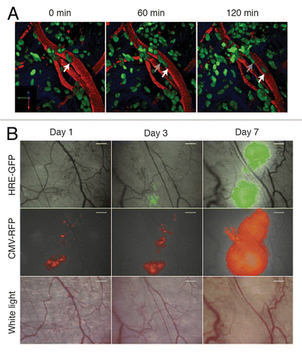
Figure 2. Most frequently used imaging windows in cancer research. (A) The CIW: the coverslip is glued onto the skull. The image depicts a coverslip and a metal ring. (B) The DSC: the metal frame of the window is clamped around the dorsal skinfold. Photograph shows the window chamber, retaining nuts and glass window. Scale bar, 1 cm. Photograph reprinted fromCitation39 by permission from Macmillan Publishers Ltd: Nature Protocols. (C) The MIW: a purse-string suture tightens the skin around the MIW. The photograph depicts the titanium MIW. . (D) The AIW: a purse-string suture secures the AIW onto the abdominal wall and skin. The photograph depicts the titanium AIW with the groove in the side and is adapted from.Citation32 All cartoons fromCitation32 Reprinted with permission from AAAS.
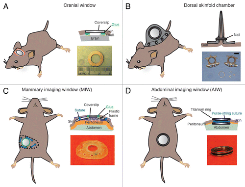
Table 1. Window surgery equipment
Table 2. Troubleshooting imaging window surgeries
Table 3. Clinical parameters of distress
Figure 3. CIW surgery. (A) The mouse head is shaved and fixed on a stereotaxic frame. (B) The skin is cut in a circular manner around the skull and the periosteum is scraped. (C) 5mm diameter circle is drawn around the region of interest. (D) A groove is drilled around the region of interest. (E) Cold cortex buffer is applied during drilling. (F) The craniotomy is lifted under a drop of saline exposing the brain. (G) The brain is covered with silicone oil and sealed with a 6 mm diameter coverslip. (H) Dental cement is applied to cover exposed skull and a stainless steel ring is glued parallel to the coverslip. (I) The mouse is injected with 100 μg/kg of buprenorphine and allowed to recover on a heating pad.
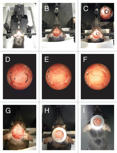
Table 4. Troubleshooting imaging
Figure 4. Purse-string suture fixes the AIW into the animal. (A) A purse-string suture is placed. The suture is first placed through skin and then through opposing abdominal wall. The outer loops (OL) are not tightened, whereas the inner loops (IL) are. (B) Once the purse-string suture is placed it should contain 4 or 5 outer loops. (C) The AIW is glued to the organ (liver [L]) and the skin and abdominal wall are placed within the window groove. (D) The sutures are tightened to secure the AIW in place. All pictures adapted fromCitation31 by permission from Macmillan Publishers Ltd: Nature Protocols.
![Figure 4. Purse-string suture fixes the AIW into the animal. (A) A purse-string suture is placed. The suture is first placed through skin and then through opposing abdominal wall. The outer loops (OL) are not tightened, whereas the inner loops (IL) are. (B) Once the purse-string suture is placed it should contain 4 or 5 outer loops. (C) The AIW is glued to the organ (liver [L]) and the skin and abdominal wall are placed within the window groove. (D) The sutures are tightened to secure the AIW in place. All pictures adapted fromCitation31 by permission from Macmillan Publishers Ltd: Nature Protocols.](/cms/asset/8c140f2f-52c5-434d-a347-3d183d5ff1e2/kinv_a_10929917_f0004.gif)
Figure 5. Fixation of the window during imaging. (A) A custom-designed imaging box that can be mounted on top of the stage of an inverted microscope. Reprinted fromCitation31 by permission from Macmillan Publishers Ltd: Nature Protocols. (B) The imaging box shown from below. MIW is fixed in the center hole. (C) Inserts for the imaging box. A CIW window insert and an AIW insert are shown. (E) An imaging mount that can be used to fix a DSC onto a microscope stage. The three smaller holes allow the bolts on the window chamber to pass through and fix it, and the large hole permits imaging through the window. Scale bar, 1 cm. reprinted fromCitation39 by permission from Mark W. Dewhirst and Macmillan Publishers Ltd: Nature Protocols. (F) An inverted Leica SP5 microscope with a dark climate chamber surrounding the stage is shown. Reprinted fromCitation31 by permission from Macmillan Publishers Ltd: Nature Protocols.
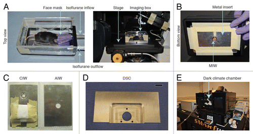
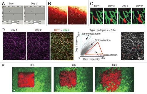
Figure 6. Retracing of the exact same field of view over multiple days. (A) A microscope with a motorized stage allows storage of regions of interest that can be imported at request. Reprinted fromCitation12 by permission from Macmillan Publishers Ltd: Nature. (B) The numbered grids on a gridded cover glass can be visualized using reflection microscopy. The numbers can be used to retrace the same position over multiple days. Reprinted fromCitation31 by permission from Macmillan Publishers Ltd: Nature. (C) The vasculature can be used as reference point to retrace a field of view. The microvasculature (green) is imaged through a cranial imaging window. Intravascular proliferation of injected PC14-PE6 lung carcinoma cells (red) can be seen from day 3 onwards. Images reprinted fromCitation22 by permission from Macmillan Publishers Ltd: Nature Medicine. (D) In the liver the type I collagen network (purple) visualized by second harmonic generation imaging was used to retrace the same position of multiple days. To show retracing, the images of day 1 and day 2 were given a false color (red or green respectively) and were merged. Yellow indicates colocalization. To quantify the amount of colocalization, the pixel intensity values of day 1 and day 2 in the merged image were displayed in a scatter plot. The Pearson correlation coefficient (r) was calculated and the non-colocalizing pixels were displayed in falce colors (red and blue) on top of the day 1 image displayed in gray. Scale bar, 20 µm. Image fromCitation32 Reprinted with permission from AAAS. (E) Dendra2 labeled tumor cells (green are non-photoswitched cells and red are photoswitched cells) imaged through a mammary imaging window. Images were taken 0, 6 and 24 h after photoswitching. Scale bar, 30 µm. Images reprinted from byCitation29 permission from Macmillan Publishers Ltd: Nature Methods.