

Figures & data
Figure 1. Generation and testing of the MIP1-CreERT transgenic mouse. (A) Transgenic construct containing MIP promoter, CreER gene and hGH and agarose gel with genotyping products from negative (1,2,5,7) and positive (3,4,6) founder lines, as well as control DNA (8); Expression of β-galactosidase in the pancreas of ROSA-LacZ:MIP1-CreERT[Tmx]: (B) Frozen sections of the pancreas from ROSA-LacZ:MIP1-CreERT mice injected or not with tamoxifen, were stained for β-galactosidase activity (blue staining); (C) frozen sections of pancreas from ROSA-LacZ:MIP1-CreERT [Tmx] mice (injected with tamoxifen) were stained for β-galactosidase (GREEN), insulin (BLUE) and glucagon (RED) antibodies. Note almost complete overlap of β-galactosidase staining and insulin staining, and the absence of such overlap of β-galactosidase with glucagon staining. Corresponding image of a control islet ROSA-LacZ:MIP1-CreERT[oil] staining for β-galactosidase, insulin and glucagon shows lack of β-galactosidase staining in the islet; (D) MIP1-CreERT transgene expression is specific for pancreatic islets: RT-PCR amplification of Cre in cDNA obtained from various tissues of MIP1-CreERT mice. Densitometry of PCR fragment in an EtBr stained gel (n = 3). Control – actin (not shown).
![Figure 1. Generation and testing of the MIP1-CreERT transgenic mouse. (A) Transgenic construct containing MIP promoter, CreER gene and hGH and agarose gel with genotyping products from negative (1,2,5,7) and positive (3,4,6) founder lines, as well as control DNA (8); Expression of β-galactosidase in the pancreas of ROSA-LacZ:MIP1-CreERT[Tmx]: (B) Frozen sections of the pancreas from ROSA-LacZ:MIP1-CreERT mice injected or not with tamoxifen, were stained for β-galactosidase activity (blue staining); (C) frozen sections of pancreas from ROSA-LacZ:MIP1-CreERT [Tmx] mice (injected with tamoxifen) were stained for β-galactosidase (GREEN), insulin (BLUE) and glucagon (RED) antibodies. Note almost complete overlap of β-galactosidase staining and insulin staining, and the absence of such overlap of β-galactosidase with glucagon staining. Corresponding image of a control islet ROSA-LacZ:MIP1-CreERT[oil] staining for β-galactosidase, insulin and glucagon shows lack of β-galactosidase staining in the islet; (D) MIP1-CreERT transgene expression is specific for pancreatic islets: RT-PCR amplification of Cre in cDNA obtained from various tissues of MIP1-CreERT mice. Densitometry of PCR fragment in an EtBr stained gel (n = 3). Control – actin (not shown).](/cms/asset/e0e4fef0-afac-4f20-887d-5aacd0e4ca89/kisl_a_10927685_f0001.gif)
Figure 2. Calcium responses in ROSALacZ:MIP1-CreERT islets. Representative traces show changes in Fura 2 fluorescence, which reflects changes in intracellular calcium in isolated mouse islets: (A) ROSA-LacZ:MIP1-CreERT[oil] males; (B) ROSA-LacZ:MIP1-CreERT[Tmx] males. Period of Ca2+ oscillation in islets was 6.7 ± 1.8 min for [Tmx]-injected and 5.9 ± 1.6 min for [oil]-injected; n = 20 islets in each group; differences in Ca2+ oscillations were not statistically significant P = 0.67). (C) - islets from (A and B) stained for β-galactosidase activity (blue color) to show minimal leakiness of Cre activity in control (ARROW) compared with Tmx-treated mice.
![Figure 2. Calcium responses in ROSALacZ:MIP1-CreERT islets. Representative traces show changes in Fura 2 fluorescence, which reflects changes in intracellular calcium in isolated mouse islets: (A) ROSA-LacZ:MIP1-CreERT[oil] males; (B) ROSA-LacZ:MIP1-CreERT[Tmx] males. Period of Ca2+ oscillation in islets was 6.7 ± 1.8 min for [Tmx]-injected and 5.9 ± 1.6 min for [oil]-injected; n = 20 islets in each group; differences in Ca2+ oscillations were not statistically significant P = 0.67). (C) - islets from (A and B) stained for β-galactosidase activity (blue color) to show minimal leakiness of Cre activity in control (ARROW) compared with Tmx-treated mice.](/cms/asset/53b8f2c3-d157-4e33-be3e-9c7283493a1f/kisl_a_10927685_f0002.gif)
Figure 3. Islet structure and physiological responses in transgenic MIP1-CreERT mice are preserved. Paraffin sections of pancreas were stained with anti-insulin (RED) and anti-glucagon (GREEN) antibodies using double immunofluorescence: (A) female non-transgenic; (B) female MIP1-CreERT+/−; (C) male non-transgenic; (D) male MIP1-CreERT+/−. Nuclei are stained with DAPI (BLUE); size bar in indicates 100µm for all panelsc (A-D). IPGTT performed on MIP1-CreERT+/− and non-transgenic littermate controls at 12 wk of age: (E) males and (F) females from each genotype were recorded separately. Each group consisted of 5–7 mice.
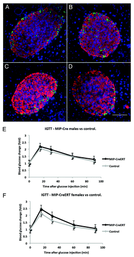
Figure 4. Tamoxifen [Tmx] injections do not alter glucose tolerance in double-transgenic ROSA-LacZ:MIP1-CreERT mice. IPGTT in 16 wk old ROSA26/LacZ:MIP1-CreERT males and females, before injection with tamoxifen and one week after a round of 5 injections (100mg/kg/day). Each group consisted of 5–7 mice.
![Figure 4. Tamoxifen [Tmx] injections do not alter glucose tolerance in double-transgenic ROSA-LacZ:MIP1-CreERT mice. IPGTT in 16 wk old ROSA26/LacZ:MIP1-CreERT males and females, before injection with tamoxifen and one week after a round of 5 injections (100mg/kg/day). Each group consisted of 5–7 mice.](/cms/asset/a2599f60-b1a2-4843-88f4-10178e2e6d94/kisl_a_10927685_f0004.gif)
Figure 5. Deleting of Ctnnb1 gene in pancreatic β-cells (βCtnnb1KO) leads to elimination of β-catenin from insulin positive cells in islets of Langerhans. Pancreatic sections stained for - β-catenin (GREEN; A, D), insulin (RED; B, E) and glucagon (BLUE, C, F). Mouse samples: (A, B, C) control (Ctnnb1fl/fl[Tmx]); (D, E, F) double-transgenic Ctnnb1KO, (Ctnnb1fl/fl:MIP1-CreERT[Tmx]). Note the decreased β-catenin staining in the islet area of Ctnnb1KO pancreas despite the preserved β–catenin staining of surrounding acinar tissue. The graph under the images represents intensity of staining along the white line shown in the β-catenin-stained panels. Note the decreased level of β-catenin intensity under the area of insulin staining indicated by red bars.
![Figure 5. Deleting of Ctnnb1 gene in pancreatic β-cells (βCtnnb1KO) leads to elimination of β-catenin from insulin positive cells in islets of Langerhans. Pancreatic sections stained for - β-catenin (GREEN; A, D), insulin (RED; B, E) and glucagon (BLUE, C, F). Mouse samples: (A, B, C) control (Ctnnb1fl/fl[Tmx]); (D, E, F) double-transgenic Ctnnb1KO, (Ctnnb1fl/fl:MIP1-CreERT[Tmx]). Note the decreased β-catenin staining in the islet area of Ctnnb1KO pancreas despite the preserved β–catenin staining of surrounding acinar tissue. The graph under the images represents intensity of staining along the white line shown in the β-catenin-stained panels. Note the decreased level of β-catenin intensity under the area of insulin staining indicated by red bars.](/cms/asset/b1747381-eea5-45eb-8262-4d463b5049bb/kisl_a_10927685_f0005.gif)
Figure 6. Effects of β-cell specific deletion Ctnnb1KO in mice include decreased expression of β-catenin in islets. Primary pancreatic islets purified from Ctnnb1KO,Ctnnb1fl/fl:MIP1-CreERT[Tmx] double-transgenic mice, and controlCtnnb1fl/fl[Tmx] mice, were stained for insulin (GREEN) and β-catenin (RED).
![Figure 6. Effects of β-cell specific deletion Ctnnb1KO in mice include decreased expression of β-catenin in islets. Primary pancreatic islets purified from Ctnnb1KO,Ctnnb1fl/fl:MIP1-CreERT[Tmx] double-transgenic mice, and controlCtnnb1fl/fl[Tmx] mice, were stained for insulin (GREEN) and β-catenin (RED).](/cms/asset/538a940f-d14e-4e22-a50b-3eb2f29c1b9e/kisl_a_10927685_f0006.gif)
Figure 7. Effects of β-cell-specific Ctnnb1 deletion. (A) IPGTT performed 4 wk after TMX injection in male and female double-transgenic Ctnnb1KO, (Ctnnb1fl/fl:MIP1-CreERT[Tmx]) as well as control,Ctnnb1fl/fl[Tmx], show no statistically significant change (n = 5 in each group). (B) Insulin secretion stimulation index (amount of insulin secreted in KRB 16.7mM glucose divided by that secreted in KRB 2.8mM glucose) is unchanged in double-transgenic Ctnnb1KO compared with control littermates. No statistically significant differences between Ctnnb1KO and control mice were detected (n = 5 in each group). (C) western blots of islet extracts from Ctnnb1KO mice and controls stained with β-catenin and actin antibodies show depletion of 90 Kd β-catenin band in the brief exposure of the western blot (TOP PANEL), while longer exposure of the same blot (LOWER PANEL) shows the appearance of additional 50Kd band, as mentioned in the text (representative of 3 independent experiments).
![Figure 7. Effects of β-cell-specific Ctnnb1 deletion. (A) IPGTT performed 4 wk after TMX injection in male and female double-transgenic Ctnnb1KO, (Ctnnb1fl/fl:MIP1-CreERT[Tmx]) as well as control,Ctnnb1fl/fl[Tmx], show no statistically significant change (n = 5 in each group). (B) Insulin secretion stimulation index (amount of insulin secreted in KRB 16.7mM glucose divided by that secreted in KRB 2.8mM glucose) is unchanged in double-transgenic Ctnnb1KO compared with control littermates. No statistically significant differences between Ctnnb1KO and control mice were detected (n = 5 in each group). (C) western blots of islet extracts from Ctnnb1KO mice and controls stained with β-catenin and actin antibodies show depletion of 90 Kd β-catenin band in the brief exposure of the western blot (TOP PANEL), while longer exposure of the same blot (LOWER PANEL) shows the appearance of additional 50Kd band, as mentioned in the text (representative of 3 independent experiments).](/cms/asset/4520ccac-40e8-4573-bd57-d4ff4f296520/kisl_a_10927685_f0007.gif)

