804
Views
35
CrossRef citations to date
0
Altmetric
Report

Development and characterization of monoclonal antibodies against Rift Valley fever virus nucleocapsid protein generated by DNA immunization

, , , , , & show all
Pages 275-284 | Received 23 Dec 2009, Accepted 02 Mar 2010, Published online: 01 May 2010

Figures & data

Figure 1 Expression and detection of nucleoprotein N. (A) of recombinant Trx (1) and Trx-N fusion protein (2) in E. coli cells as detected by Coomassie staining of Ni-NTA column purified bacterial extracts, or by anti-RVFV rabbit (3) or sheep (4) hyperinmune sera. (B) Inmunoprecipitation of BHK21 cell supernatants (S) and cell lysates (CL) with a rabbit anti-RVFV polyclonal serum. (1) MP12 infected cells, (2) mock infected cells, (3) pCMV transfected cells and (4) pCMV-N transfected cell extracts. (C) Inmunoprecipitation of supernatant (S) and cell lysates (CL) of MP12 infected BHK21 cells using serum from mice immunized with pCMV-N (2 and 3) or sheep anti-RVFV hyperinmune serum (1). Mr: relative molecular mass in kilodaltons.
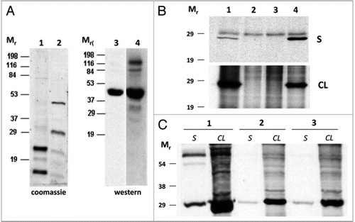
Figure 1 Expression and detection of nucleoprotein N. (A) of recombinant Trx (1) and Trx-N fusion protein (2) in E. coli cells as detected by Coomassie staining of Ni-NTA column purified bacterial extracts, or by anti-RVFV rabbit (3) or sheep (4) hyperinmune sera. (B) Inmunoprecipitation of BHK21 cell supernatants (S) and cell lysates (CL) with a rabbit anti-RVFV polyclonal serum. (1) MP12 infected cells, (2) mock infected cells, (3) pCMV transfected cells and (4) pCMV-N transfected cell extracts. (C) Inmunoprecipitation of supernatant (S) and cell lysates (CL) of MP12 infected BHK21 cells using serum from mice immunized with pCMV-N (2 and 3) or sheep anti-RVFV hyperinmune serum (1). Mr: relative molecular mass in kilodaltons.

Figure 2 Detection of nucleoprotein N in infected cells. (A) Western blot of MP12 infected cellular extracts and inmunoblotting with six different mAb supernatants. C+: mouse hyperimmune anti-RVFV serum; C-: preinmmune mouse serum. Relative molecular mass (Mr) is given in kilodaltons. (B) Immunofluorescence of MP12 and South African RVFV strain AR20368 infected Vero cells using the six mAb supernatants tested in (A). (C) Immunoprecipitation of nucleoprotein N expressed in BHK-21 cells infected with ZH548-MP12 or with South African virulent isolates, using mAbs D7D8 (1) and D7E8 (2). A polyclonal mouse anti-RVFV serum (3) was used as positive control. Detection of the immunoprecipitated antigen was performed by western blot using the purified D9D11 mAb conjugated to peroxidase.
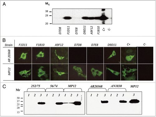
Figure 2 Detection of nucleoprotein N in infected cells. (A) Western blot of MP12 infected cellular extracts and inmunoblotting with six different mAb supernatants. C+: mouse hyperimmune anti-RVFV serum; C-: preinmmune mouse serum. Relative molecular mass (Mr) is given in kilodaltons. (B) Immunofluorescence of MP12 and South African RVFV strain AR20368 infected Vero cells using the six mAb supernatants tested in (A). (C) Immunoprecipitation of nucleoprotein N expressed in BHK-21 cells infected with ZH548-MP12 or with South African virulent isolates, using mAbs D7D8 (1) and D7E8 (2). A polyclonal mouse anti-RVFV serum (3) was used as positive control. Detection of the immunoprecipitated antigen was performed by western blot using the purified D9D11 mAb conjugated to peroxidase.

Figure 3 (A) Conservation plot of the 1–60 (upper) and 121–180 (lower) amino acid regions of the RVFV nucleoprotein N from several Egyptian lineage isolates and the South African strains used in this study. Only the regions of the primary sequence in which amino acid changes were found are showed. (B) Immunofluorescence of transfected BHK-21 cells with pCMV-NAR20368 and pCMV-NMP12 constructs. Expression of N protein was detected with the indicated mAbs.
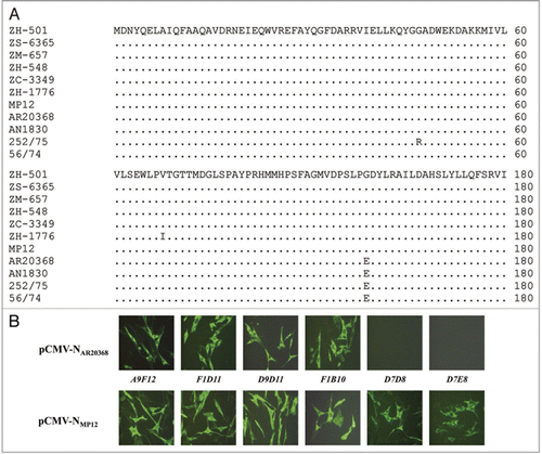
Figure 3 (A) Conservation plot of the 1–60 (upper) and 121–180 (lower) amino acid regions of the RVFV nucleoprotein N from several Egyptian lineage isolates and the South African strains used in this study. Only the regions of the primary sequence in which amino acid changes were found are showed. (B) Immunofluorescence of transfected BHK-21 cells with pCMV-NAR20368 and pCMV-NMP12 constructs. Expression of N protein was detected with the indicated mAbs.

Figure 4 Competitive ELISA assay (C-ELISA). Inhibition percentages of six mAb supernatants plotted against different dilutions of the competing sera. OVI reference anti-RVFV pooled sheep sera (♦); sera from experimentally MP12 vaccinated sheep (▪); hyperinmune rabbit anti-RVFV serum (x) and negative pooled sheep sera (▲). Data represent the mean ± SD from three replicate experiments.
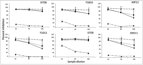
Figure 4 Competitive ELISA assay (C-ELISA). Inhibition percentages of six mAb supernatants plotted against different dilutions of the competing sera. OVI reference anti-RVFV pooled sheep sera (♦); sera from experimentally MP12 vaccinated sheep (▪); hyperinmune rabbit anti-RVFV serum (x) and negative pooled sheep sera (▲). Data represent the mean ± SD from three replicate experiments.

Figure 5 (A) Discrimination of RVFV positive and negative serum samples in C-ELISA. the percentages of inhibition obtained for each mAb were grouped in classes and plotted against the respective percentage of confirmed RVFV negative (solid bars) and positive (open bars) serum samples.
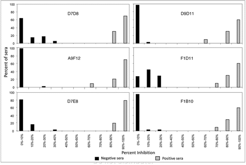
Figure 5 (A) Discrimination of RVFV positive and negative serum samples in C-ELISA. the percentages of inhibition obtained for each mAb were grouped in classes and plotted against the respective percentage of confirmed RVFV negative (solid bars) and positive (open bars) serum samples.

Figure 6 (A) Performance of mAb F1D11 and A9F12 as capture antibodies in ELISA. Positive (experimentally vaccinated sheep, shaded bars and rabbit anti-RVFV hyperinmune serum, open bar) and negative sheep sera (1/120 dilution) were tested in ELISA for their ability to bind to mAb captured viral antigen from infected cell culture supernatants. An anti-sheep HRP-conjugated (diluted to 1/6,000) was used for detection of the inmunocomplexes. (B) Competitive using either purified A9F12 mAb or rabbit anti-RVFV serum as capture antibodies. experimental sera from vaccinated sheep were used to compete the binding of A9F12-HRP labelled antibody.
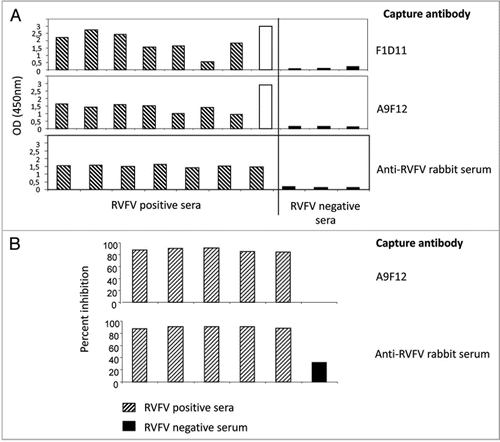
Figure 6 (A) Performance of mAb F1D11 and A9F12 as capture antibodies in ELISA. Positive (experimentally vaccinated sheep, shaded bars and rabbit anti-RVFV hyperinmune serum, open bar) and negative sheep sera (1/120 dilution) were tested in ELISA for their ability to bind to mAb captured viral antigen from infected cell culture supernatants. An anti-sheep HRP-conjugated (diluted to 1/6,000) was used for detection of the inmunocomplexes. (B) Competitive using either purified A9F12 mAb or rabbit anti-RVFV serum as capture antibodies. experimental sera from vaccinated sheep were used to compete the binding of A9F12-HRP labelled antibody.

Table 1 Characterization of mAbs anti-RVFV nucleoprotein

Table 2 Percent of binding inhibition of labelled antibodies in competitive ELISA

Reprints and Corporate Permissions

Please note: Selecting permissions does not provide access to the full text of the article, please see our help page How do I view content?

To request a reprint or corporate permissions for this article, please click on the relevant link below:

Academic Permissions

Please note: Selecting permissions does not provide access to the full text of the article, please see our help page How do I view content?

Obtain permissions instantly via Rightslink by clicking on the button below:

If you are unable to obtain permissions via Rightslink, please complete and submit this Permissions form. For more information, please visit our Permissions help page.