Figures & data
Figure 1 Neutralization of IL-21 dependent proliferation of BaF3 cells expressing IL-21R. Human IL-21R-transfected BaF3 cells (A) and murine IL-21R-transfected BaF3 cells (B) were treated with antibodies and human or murine IL-21, respectively, for 48 hours and their proliferation measured by CellTiter Glo. Antibodies tested were the parental 18A5 (solid circles), Ab-01 (open squares), Ab-02 (open triangles), Ab-03 (open circles), control IgG (X), and the human or murine IL-21R-Fc (solid squares).
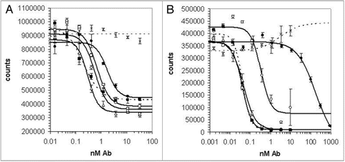
Figure 2 CDR3 sequences of optimized anti-IL-21R antibodies. The frequencies of different amino acids at each position of heavy chain CDR3 (A) or light chain CDR3 (B) in the 25 selected sequences with the highest potency are indicated by the height of the colored sections of each bar. The sequence of the 18A5 at the equivalent position is indicated below the graph. Blue lines show the sequences randomized in libraries VH-A, VH-B, VL-A and VL-B. CDR3 sequences of the clones chosen for detailed analysis are shown in (C).
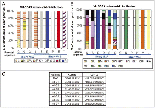
Figure 3 Serum concentration-time profiles of Ab-01 and Ab-02 following a single 10 mg/kg dose to MRL-Faslpr mice or Sprague-Dawley rats. Ab-01 (filled circles), Ab-02 (open circles), or an isotype control anti-human IL-13 antibody (open triangles in (B) only) were administered to ∼12-week old MRL-Faslpr mice (A, IPdose) or Sprague-Dawley rats (B, IV dose) and test article concentrations in serum were determined by ELISA, as described in the text. Individual concentration values below the limit of quantitation (LOQ of 33.4–66.8 ng/mL for anti-IL-21R antibodies and 132 ng/mL for an isotype control antibody) were treated as zero for calculations of the mean and standard deviation. Non-serial sampling design was used for mice (n = 6–9 per time point) and serial sampling design was used for rats (n = 4–6 per group). Time points at which all animals showed concentration values below the LOQ are indicated by ovals.
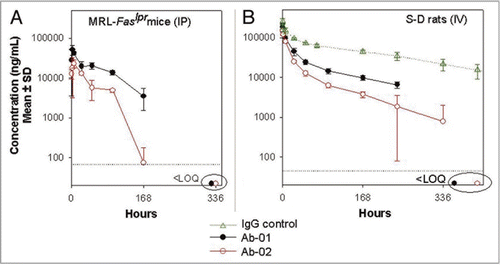
Figure 4 Serum concentration-time profiles of Ab-01 and Ab-02 following a single dose to cynomolgus monkeys. Ab-01 (filled circles in panels A and B and as indicated in panel C), Ab-02 (open circles in A and B), or an isotype control anti-human IL-13 antibody (open triangles in A) were administered to cynomolgus monkeys at indicated dose levels and routes (n = 3–4 per group), and test article concentrations in serum were determined by ELISA, as described in the text. Mean (A and B) or individual animal (C) concentrations are shown. Individual concentration values below the limit of quantitation (LOQ of 30 ng/mL for anti-IL-21R antibodies and 4 ng/mL for an isotype control) were treated as zero for calculations of the mean and standard deviation. Time points at which all (A) or specified (C) animals showed concentration values below the LOQ are indicated by ovals.
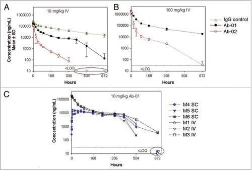
Figure 5 Median titer of anti-dsDNA antibodies in MRL-Faslpr mice treated with anti-IL-21R antibodies. Twelve week old MRL-Faslpr mice were administered Ab-01 (open triangles), Ab-02 (open circles), saline (filled diamonds), or an isotype control anti-human IL-13 antibody (filled squares) at 400 µg/mouse (10 mg/kg), 3x/week IP for 10 weeks and sacrificed at 3 days after the last dose. Serum was collected prior to dosing (week 0) and then at 2, 4, 6, 8 and 10 weeks post-dose, and mouse anti-dsDNA antibodies were monitored by ELISA, as described in the text. Limit of detection was 1,000 units/mL. Individual data values that were below the lower limit of detection were treated as 1,000 units/mL for calculations of the group median titers. *p < 0.05, based on a Mann-Whitney test using baseline, the saline group and the isotype control IgG group as a control.
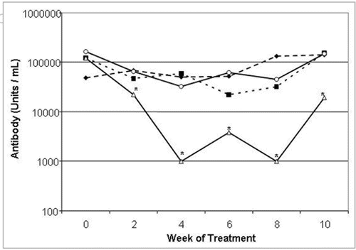
Figure 6 IgG deposits in the kidneys of MRL-Faslpr mice treated with anti-IL-21R antibodies. Twelve week old MRL-Faslpr mice were administered Ab-01, Ab-02, saline, or an isotype control anti-human IL-13 antibody at 400 µg/mouse (10 mg/kg), 3x/week IP for 10 weeks and sacrificed at 3 days after the last dose. IgG deposits in the kidney sections were assessed by immunohistochemistry and scored blindly on a scale of 0 (none) to 4 (severe) at the 10 week time point. Group mean scores (line) and individual animal scores (A), as wells as representative pictures (B) for each group are shown. *p < 0.05, based on a Mann-Whitney test using the saline and the isotype control IgG group as a control.
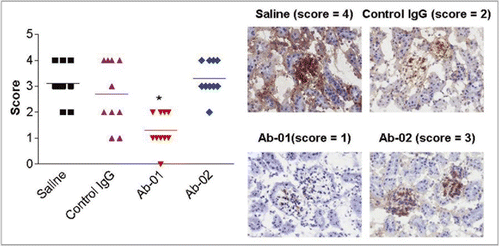
Table 1 Binding and neutralization properties of anti-IL-21 receptor antibodies
Table 2 Mean (±SD) PK parameters of Ab-01 and Ab-02 in mice, rats and monkeys