2,299
Views
24
CrossRef citations to date
0
Altmetric
Report

Suppression of innate and adaptive B cell activation pathways by antibody coengagement of FcγRIIb and CD19

, , , , &
Pages 991-999 | Received 15 Jan 2014, Accepted 09 Apr 2014, Published online: 23 Apr 2014

Figures & data

Figure 1. XmAb5871 suppresses BCR and TLR9-induced signals through phosphorylation of FcγRIIb. Purified human resting tonsillar B cells were pre-treated with media or 10 μg/ml XENP6187 or XmAb5871, respectively, for 1 h before activation with anti-Ig (2.5 μg/ml) or CpG (1 μg/ml) for 30 min. Cell lysates were western-blotted to assess phosphorylation and activation of FcγRIIb, AKT and ERK. One representative experiment from three independent ones is shown.
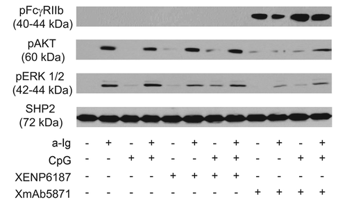
Figure 1. XmAb5871 suppresses BCR and TLR9-induced signals through phosphorylation of FcγRIIb. Purified human resting tonsillar B cells were pre-treated with media or 10 μg/ml XENP6187 or XmAb5871, respectively, for 1 h before activation with anti-Ig (2.5 μg/ml) or CpG (1 μg/ml) for 30 min. Cell lysates were western-blotted to assess phosphorylation and activation of FcγRIIb, AKT and ERK. One representative experiment from three independent ones is shown.

Figure 2. XmAb5871 inhibits while XENP6187 enhances calcium mobilization when CD19 is coligated with BCR. Fluo-4 loaded B cells were treated with either XmAb5871 or the Fc-KO XENP6187 antibodies and then stimulated with 10 μg/ml anti-Ig. Calcium flux kinetics was recorded using FACSCalibur flow cytometer and data were analyzed by FlowJo software. Data represent the average of two independent experiments.
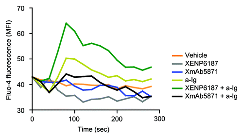
Figure 2. XmAb5871 inhibits while XENP6187 enhances calcium mobilization when CD19 is coligated with BCR. Fluo-4 loaded B cells were treated with either XmAb5871 or the Fc-KO XENP6187 antibodies and then stimulated with 10 μg/ml anti-Ig. Calcium flux kinetics was recorded using FACSCalibur flow cytometer and data were analyzed by FlowJo software. Data represent the average of two independent experiments.

Figure 3. Effect of XmAb5871 on BCR and TLR9-induced proliferation. (A) Representative flow cytometry histograms of CFSE-labeled human blood B cells stimulated with combinations of 2.5 μg/ml anti-Ig and 1 μg/ml CpG in the presence of 10 μg/ml XENP6187 or XmAb5871 for 5 d. Peaks shifted to the left represent cell populations undergoing increasing numbers of cell division. Total percentages of dividing cells are shown. (B) Percentages of dividing cells (mean ± SD) in three independent experiments. B cells were stimulated with anti-Ig, CpG ODN or with the combination of the two in the presence or absence of XENP6187 or XmAb5871. XmAb5871 significantly reduced proliferation of both CpG and anti-Ig plus CpG stimulated cells, *: P < 0.05.
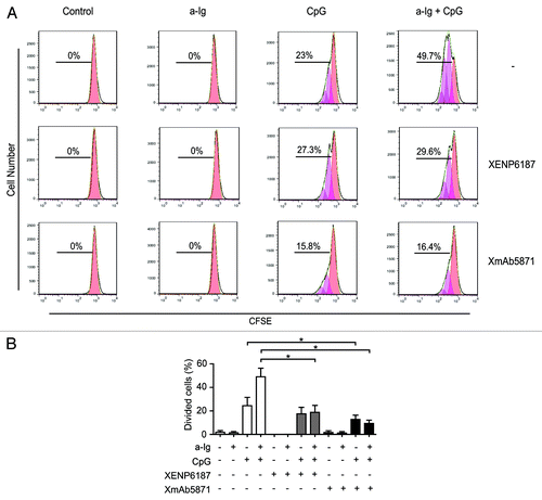
Figure 3. Effect of XmAb5871 on BCR and TLR9-induced proliferation. (A) Representative flow cytometry histograms of CFSE-labeled human blood B cells stimulated with combinations of 2.5 μg/ml anti-Ig and 1 μg/ml CpG in the presence of 10 μg/ml XENP6187 or XmAb5871 for 5 d. Peaks shifted to the left represent cell populations undergoing increasing numbers of cell division. Total percentages of dividing cells are shown. (B) Percentages of dividing cells (mean ± SD) in three independent experiments. B cells were stimulated with anti-Ig, CpG ODN or with the combination of the two in the presence or absence of XENP6187 or XmAb5871. XmAb5871 significantly reduced proliferation of both CpG and anti-Ig plus CpG stimulated cells, *: P < 0.05.

Figure 4. XmAb5871 inhibits BCR and TLR9-induced IL-6, IL-10 and TNF production by B cells. B cells cultured in 96-well plates were activated by 2.5 μg/ml anti-Ig or 1 μg/ml CpG ODN in the presence of XmAb5871 or the control antibody XENP6187. IL-6, IL-10 and TNF secreted from the culture supernatants were measured after 48 h using the Flow Cytomix bead array. Data represent the mean ± SD of four independent experiments. XmAb5871 significantly inhibits IL-6 production induced by anti-Ig and CpG, as compared with XENP6187 treated cells. *: P < 0.05.
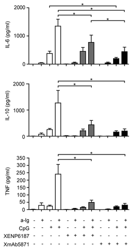
Figure 4. XmAb5871 inhibits BCR and TLR9-induced IL-6, IL-10 and TNF production by B cells. B cells cultured in 96-well plates were activated by 2.5 μg/ml anti-Ig or 1 μg/ml CpG ODN in the presence of XmAb5871 or the control antibody XENP6187. IL-6, IL-10 and TNF secreted from the culture supernatants were measured after 48 h using the Flow Cytomix bead array. Data represent the mean ± SD of four independent experiments. XmAb5871 significantly inhibits IL-6 production induced by anti-Ig and CpG, as compared with XENP6187 treated cells. *: P < 0.05.

Figure 5. The effect of XmAb5871 on total and citrullinated peptide-specific IgG production as detected by ELISPOT assay (A) Purified human B cells were cultured with anti-Ig (2.5 μg/ml) or CpG (0.5 μg/ml) for 3 d in the presence of IL-2 (50 ng/ml) and IL-10 (50 ng/ml), and XmAb5871 or XENP6187 (10 μg/ml), respectively. The number of IgG-secreting cells was evaluated by ELISPOT assay on anti-IgG-coated nitrocellulose plates. Data represent the mean ± SD of seven independent experiments. *: P < 0.05. (B) B cells were stimulated by IL-2 (10 ng/ml) and R848 (1 μg/ml) for 3 d in the presence of XENP6187 or XmAb5871 antibodies. The number of IgG-secreting cells was assessed as above. Data represent the mean ± SD of seven independent experiments. *: P < 0.05. (C) Citrullinated filaggrin peptide-specific IgG-producing B cells were tested upon activation with IL-2 (10 ng/ml) and R848 (1 μg/ml) for 3 d with or without XmAb5871. The citrullinated filaggrin peptide-specific IgG secreting cells were detected on peptide-coated nitrocellulose plates. The results for B cells from six different ACPA-positive RA patients are shown. XmAb5871 significantly inhibited the development of citrullin-containing filaggrin peptide-specific antibody-forming cells, *: P < 0.05.
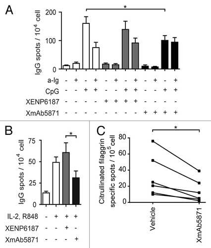
Figure 5. The effect of XmAb5871 on total and citrullinated peptide-specific IgG production as detected by ELISPOT assay (A) Purified human B cells were cultured with anti-Ig (2.5 μg/ml) or CpG (0.5 μg/ml) for 3 d in the presence of IL-2 (50 ng/ml) and IL-10 (50 ng/ml), and XmAb5871 or XENP6187 (10 μg/ml), respectively. The number of IgG-secreting cells was evaluated by ELISPOT assay on anti-IgG-coated nitrocellulose plates. Data represent the mean ± SD of seven independent experiments. *: P < 0.05. (B) B cells were stimulated by IL-2 (10 ng/ml) and R848 (1 μg/ml) for 3 d in the presence of XENP6187 or XmAb5871 antibodies. The number of IgG-secreting cells was assessed as above. Data represent the mean ± SD of seven independent experiments. *: P < 0.05. (C) Citrullinated filaggrin peptide-specific IgG-producing B cells were tested upon activation with IL-2 (10 ng/ml) and R848 (1 μg/ml) for 3 d with or without XmAb5871. The citrullinated filaggrin peptide-specific IgG secreting cells were detected on peptide-coated nitrocellulose plates. The results for B cells from six different ACPA-positive RA patients are shown. XmAb5871 significantly inhibited the development of citrullin-containing filaggrin peptide-specific antibody-forming cells, *: P < 0.05.

Reprints and Corporate Permissions

Please note: Selecting permissions does not provide access to the full text of the article, please see our help page How do I view content?

To request a reprint or corporate permissions for this article, please click on the relevant link below:

Academic Permissions

Please note: Selecting permissions does not provide access to the full text of the article, please see our help page How do I view content?

Obtain permissions instantly via Rightslink by clicking on the button below:

If you are unable to obtain permissions via Rightslink, please complete and submit this Permissions form. For more information, please visit our Permissions help page.