1,067
Views
34
CrossRef citations to date
0
Altmetric
Report

The in vitro resistance of IgG2 to proteolytic attack concurs with a comparative paucity of autoantibodies against peptide analogs of the IgG2 hinge

, , &
Pages 558-567 | Received 10 Aug 2011, Accepted 16 Sep 2011, Published online: 04 Nov 2011

Figures & data

Figure 1 Human IgG2 is resistant to cleavage by a number of physiologically-relevant proteases. (A) Purified human IgG1 was incubated with different proteases and analyzed by capillary electrophoresis under denaturing, non-reducing conditions. Specific enzymes are noted above individual lanes, and all digestions were carried out for 24 h at 37°C. The far right three lanes have purified human IgG1 standards, representing the following: Lane 9, single cleaved IgG1 (scIgG1); Lane 10, F(ab')2 fragment of IgG1; Lane 11, intact IgG1. The Fc monomer released under denaturing conditions is labeled as Fc(m). (B) Lanes 2–8 depict human IgG2 incubated with different proteases analyzed by capillary electrophoresis under denaturing, non-reducing conditions. The same standards used in (A) were run in Lanes 9–11. (C) Bar graph representation of the percent of intact IgG remaining after 24 h of proteolytic digestions. Open bars represent human IgG1 and shaded bars represent human IgG2. Bar heights correspond to the mean ± SD from four independent tests.
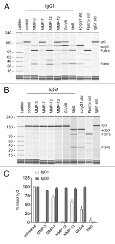
Figure 1 Human IgG2 is resistant to cleavage by a number of physiologically-relevant proteases. (A) Purified human IgG1 was incubated with different proteases and analyzed by capillary electrophoresis under denaturing, non-reducing conditions. Specific enzymes are noted above individual lanes, and all digestions were carried out for 24 h at 37°C. The far right three lanes have purified human IgG1 standards, representing the following: Lane 9, single cleaved IgG1 (scIgG1); Lane 10, F(ab')2 fragment of IgG1; Lane 11, intact IgG1. The Fc monomer released under denaturing conditions is labeled as Fc(m). (B) Lanes 2–8 depict human IgG2 incubated with different proteases analyzed by capillary electrophoresis under denaturing, non-reducing conditions. The same standards used in (A) were run in Lanes 9–11. (C) Bar graph representation of the percent of intact IgG remaining after 24 h of proteolytic digestions. Open bars represent human IgG1 and shaded bars represent human IgG2. Bar heights correspond to the mean ± SD from four independent tests.

Figure 2 Human anti-hinge autoantibodies were detected against peptide analogs of the human IgG1 hinge region but not human IgG2. (A) The sequence of human IgG1 (top) and human IgG2 (bottom) ranging from amino acids 212–241 (EU numbering). (B) ELISA binding of IgG3 autoantibodies from serum pooled from 20 healthy donors to peptide analogs of the human IgG1 and IgG2 hinge/CH2 regions. Letters on the X-axis represent the free C-termini of 14-mer peptides. (C) ELISA binding of IgG autoantibodies from serum pooled from 20 healthy donors to peptide analogs of the human IgG1 and IgG2 hinge/CH2 regions.
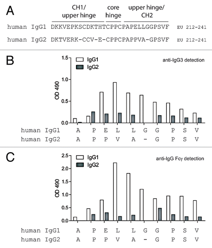
Figure 2 Human anti-hinge autoantibodies were detected against peptide analogs of the human IgG1 hinge region but not human IgG2. (A) The sequence of human IgG1 (top) and human IgG2 (bottom) ranging from amino acids 212–241 (EU numbering). (B) ELISA binding of IgG3 autoantibodies from serum pooled from 20 healthy donors to peptide analogs of the human IgG1 and IgG2 hinge/CH2 regions. Letters on the X-axis represent the free C-termini of 14-mer peptides. (C) ELISA binding of IgG autoantibodies from serum pooled from 20 healthy donors to peptide analogs of the human IgG1 and IgG2 hinge/CH2 regions.

Figure 3 Whole blood ADCC and CDC using human IgG1 and IgG2 anti-CD20 intact mAbs and F(ab')2 fragments generated with IdeS. (A) Human donor one whole blood ADCC activity against WIL2-S cells using intact IgG1 anti-CD20 (black circles), IgG1 F(ab')2 of anti-CD20 (red squares), IgG2 anti-CD20 (blue up triangle), IgG2 F(ab')2 of anti-CD20 (green down triangle), and an IgG1 isotype control (black diamond) (n = 2). (B) Human donor two whole blood ADCC activity against WIL2-S cells (n = 2). (C) Cell-based CDC against WIL2-S cells containing 50% serum from human donor one (n = 2). (D) Cell-based CDC against WIL2-S cells containing 50% serum from human donor two (n = 2).
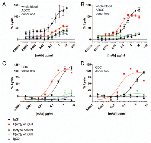
Figure 3 Whole blood ADCC and CDC using human IgG1 and IgG2 anti-CD20 intact mAbs and F(ab')2 fragments generated with IdeS. (A) Human donor one whole blood ADCC activity against WIL2-S cells using intact IgG1 anti-CD20 (black circles), IgG1 F(ab')2 of anti-CD20 (red squares), IgG2 anti-CD20 (blue up triangle), IgG2 F(ab')2 of anti-CD20 (green down triangle), and an IgG1 isotype control (black diamond) (n = 2). (B) Human donor two whole blood ADCC activity against WIL2-S cells (n = 2). (C) Cell-based CDC against WIL2-S cells containing 50% serum from human donor one (n = 2). (D) Cell-based CDC against WIL2-S cells containing 50% serum from human donor two (n = 2).

Figure 4 ADCP using human IgG1 and IgG2 anti-CD142 intact mAbs and F(ab')2 fragments generated with IdeS. (A) Representative flow cytometry dot plots depicting GFP-expressing MDA-MB-231 cells and human macrophages detected with anti-CD11b and anti-CD14 mAbs conjugated to Alexa Fluor 647. The left plot depicts cells incubated in the absence of anti-CD142 mAbs and the plot on the right depicts cells incubated with 9 µg/ml human IgG1 intact anti-CD142 mAb. (B) Representative fluorescent microscopy images of macrophages and GFP-expressing MDA-MB-231 cells in the absence of mAb (left panel) and incubated with 9 µg/ml IgG1 intact anti-CD142 (right part). Macrophages were detected with anti-CD11b and anti-CD14 antibodies conjugated to Alexa Fluor 568 (red), whereas the green indicates GFP-expressing MDA-MB-231 cells. The white arrows indicate macrophages that had phagocytosed MDA-MB-231 cells. (C) Human macrophage-mediated ADCP activity against GFP-expressing cells using intact IgG1 anti-CD142 (black circles), IgG1 F(ab')2 of anti-CD142 (red squares), IgG2 anti-CD142 (blue up triangle), and IgG2 F(ab')2 of anti-CD142 (green down triangle) in the absence of human serum. (D) ADCP activity against GFP-expressing cells in the presence of 30% human serum from donor one (n = 2). (E) ADCP activity against GFP-expressing cells in the presence of 30% human serum from donor two (n = 2).
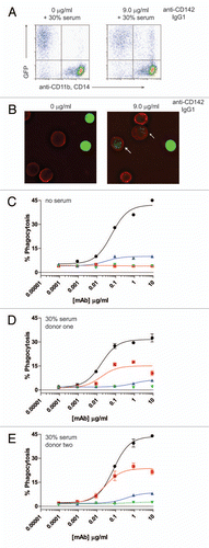
Figure 4 ADCP using human IgG1 and IgG2 anti-CD142 intact mAbs and F(ab')2 fragments generated with IdeS. (A) Representative flow cytometry dot plots depicting GFP-expressing MDA-MB-231 cells and human macrophages detected with anti-CD11b and anti-CD14 mAbs conjugated to Alexa Fluor 647. The left plot depicts cells incubated in the absence of anti-CD142 mAbs and the plot on the right depicts cells incubated with 9 µg/ml human IgG1 intact anti-CD142 mAb. (B) Representative fluorescent microscopy images of macrophages and GFP-expressing MDA-MB-231 cells in the absence of mAb (left panel) and incubated with 9 µg/ml IgG1 intact anti-CD142 (right part). Macrophages were detected with anti-CD11b and anti-CD14 antibodies conjugated to Alexa Fluor 568 (red), whereas the green indicates GFP-expressing MDA-MB-231 cells. The white arrows indicate macrophages that had phagocytosed MDA-MB-231 cells. (C) Human macrophage-mediated ADCP activity against GFP-expressing cells using intact IgG1 anti-CD142 (black circles), IgG1 F(ab')2 of anti-CD142 (red squares), IgG2 anti-CD142 (blue up triangle), and IgG2 F(ab')2 of anti-CD142 (green down triangle) in the absence of human serum. (D) ADCP activity against GFP-expressing cells in the presence of 30% human serum from donor one (n = 2). (E) ADCP activity against GFP-expressing cells in the presence of 30% human serum from donor two (n = 2).
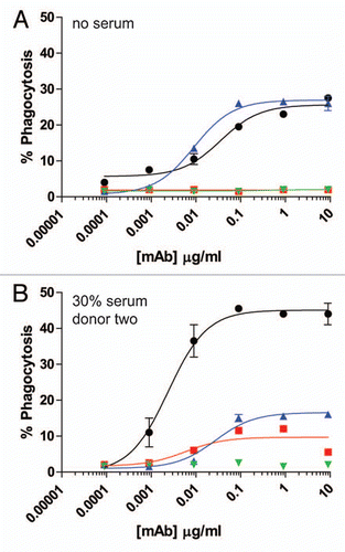
Figure 5 ADCP using human IgG1 and IgG2 anti-EGFR intact mAbs and F(ab')2 fragments generated with IdeS. (A) Human macrophage-mediated ADCP activity against GFP-expressing cells using intact IgG1 anti-EGFR (black circles), IgG1 F(ab')2 of anti-EGFR (red squares), IgG2 anti-EGFR (blue up triangle), and IgG2 F(ab')2 of anti-EGFR (green down triangle) in the absence of human serum (n = 2). (B) ADCP activity against GFP-expressing cells in the presence of 30% human serum from donor two (n = 2).

Figure 5 ADCP using human IgG1 and IgG2 anti-EGFR intact mAbs and F(ab')2 fragments generated with IdeS. (A) Human macrophage-mediated ADCP activity against GFP-expressing cells using intact IgG1 anti-EGFR (black circles), IgG1 F(ab')2 of anti-EGFR (red squares), IgG2 anti-EGFR (blue up triangle), and IgG2 F(ab')2 of anti-EGFR (green down triangle) in the absence of human serum (n = 2). (B) ADCP activity against GFP-expressing cells in the presence of 30% human serum from donor two (n = 2).

Reprints and Corporate Permissions

Please note: Selecting permissions does not provide access to the full text of the article, please see our help page How do I view content?

To request a reprint or corporate permissions for this article, please click on the relevant link below:

Academic Permissions

Please note: Selecting permissions does not provide access to the full text of the article, please see our help page How do I view content?

Obtain permissions instantly via Rightslink by clicking on the button below:

If you are unable to obtain permissions via Rightslink, please complete and submit this Permissions form. For more information, please visit our Permissions help page.