Figures & data
Figure 1 4–15% SDS-PAGE of murine and cRFB4 constructs under non-reducing (A) and reducing (B) conditions. Lane 1, cRFB4; lane 2, mcRFB4-P247W; lane 3, mcRFB4-I253A; lane 4, mcRFB4-H310A; lane 5, mcRFB4-H435A; lane 6, mcRFB4-AAA; lane 7, murine RFB4. This is one of three experiments.
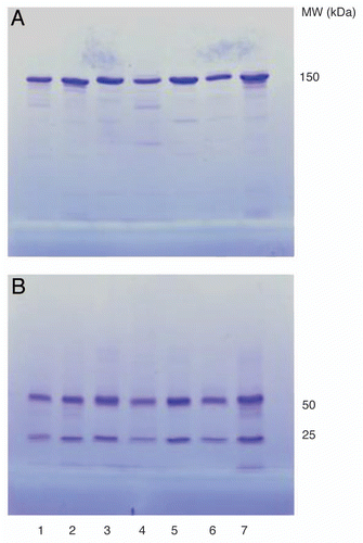
Figure 2 Analysis of purified wild-type cRFB4 and His310Ala constructs using size-exclusion chromatography. (A) cRFB4; (B) mcRFB4-H310A. This is one of three experiments.
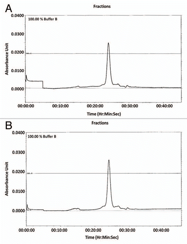
Figure 3 Autoradiograph of murine and chimeric RFB4 constructs on SDS gels. 125I-labeled mAbs were incubated for 24 h with mouse serum at 37°C and then 4–15% SDS-PAGE was performed under non-reducing (A) or reducing (B) conditions. Lane 1, murine RFB4; lane 2, cRFB4; lane 3, mcRFB4-P247W; lane 4, mcRFB4-I253A; lane 5, mcRFB4-H310A; lane 6, mcRFB4-H435A; lane 7, mcRFB4-AAA. This is one of three experiments.
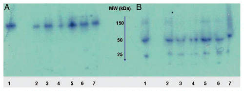
Figure 4 Analysis of the stability of cRFB4 constructs in mouse serum in vivo. (A) cRFB4: time 0 h; (B) cRFB4: time 24 h; (C) mcRFB4-H310A: time 0 h. (D) mcRFB4-H310A: time 24 h. Black line: measurement of radioactivity in cpm.; red line: measurement of the absorbance at 280 nm. IgM, immunoglobulin M; IgG, immunoglobulin G; Alb, albumin. This is one of two experiments.
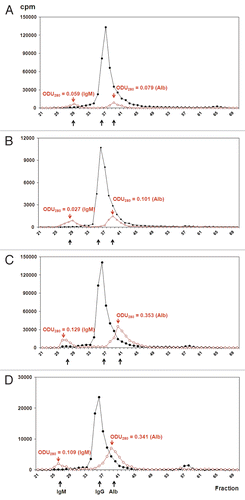
Figure 5 4–15% SDS-PAGE of murine and chimeric RFB4 ITs under non-reducing conditions. Lane 1, RFB4-rRTA; lane 2, cRFB4-rRTA; lane 3, mcRFB4-P247W-rRTA; lane 4, mcRFB4-H310A-rRTA. This is one of three experiments.
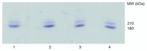
Figure 6 Antigen-binding activity of the ITs vs. the corresponding cmAbs as determined by FACS. (A) (▴) cRFB4; (●) cRFB4-rRTA; (B) (▴) mcRFB4-P247W; (●) mcRFB4-P247W-rRTA; (C) (▴) mcRFB4-H310A; (●) mcRFB4-H310A-rRTA. This is one of three experiments.
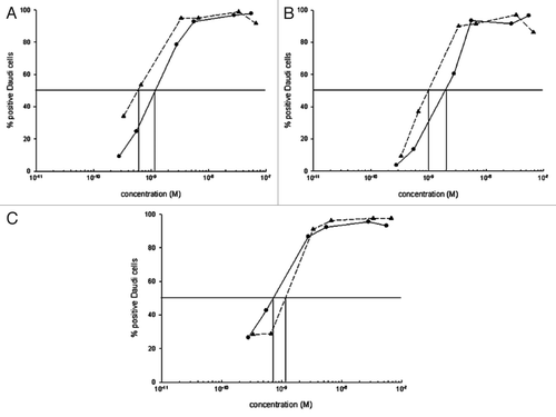
Figure 7 PVL of anti-CD22 ITs in mice. Open column, RFB4-rRTA; Gray column, cRFB4-rRTA; Black column, mcRFB4-H310A-rRTA. Murine RFB4-rRTA and cRFB4-rRTA ITs showed similar toxicity regardless the doses (p > 0.26), while at the dose of 7.5 mg/kg, mcRFB4-H310A-rRTA is significantly less toxic as compared with both murine and chimeric RFB4 ITs (p < 0.04). This is one of two experiments.
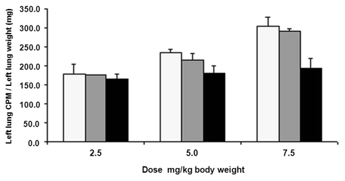
Figure 8 Changes in the body weight of BALB/c mice after treatment with various doses of ITs. (A) 2.5 mg/kg; (B) 5 mg/kg; (C) 7.5 mg/kg; (■) RFB4-rRTA; (□) cRFB4-rRTA; (▴) mcRFB4-H310A-rRTA. This is one of two experiments.
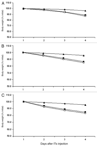
Table 1 Pharmacokinetics of cRFB4 constructs in Swiss Webster miceTable Footnotea
Table 2 Cytotoxicity of ITs on CD22+ Daudi tumor cellsTable Footnotea
Table 3 Pharmacokinetics of ITs and their corresponding mAbs in BALB/c miceTable Footnotea