Figures & data
Figure 1 Detection of native antigens in a crude parasite extract dilution from P. falciparum culture with selected monoclonal antibodies. (A) HRP2 antibodies (C1–13 and PTL3, triangles) compared with strongest HDP (marked with H, solid lines) and DHFR-TS (marked with D, open squares) antibodies, (B) DHFR-TS antibodies, (C) HDP antibodies and (D) GLURP antibodies.
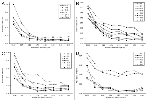
Table 1 Antibody response of mouse sera to the recombinant antigen in ELISA at different days before each (booster) immunization during the immunization procedure
Table 2 Overview and characteristics of hybridoma clones during immunization and selection process
Table 3 Dissociation constants (KD) of monoclonal antibodies as determined by ELISA
Table 4 Detection limit of crude parasite antigen in ELISA by selected antibodies compared with HRP2 antibodies
Table 5 Primer sequences for recombinant antigens and encoding nucleotide range