Figures & data
Figure 1. (A) Antitumor activity of metronomic cyclophosphamide, single low-dose cyclophosphamide, AdIL-12 and their combinations in the CT26 colorectal carcinoma model. Animals were distributed into different groups: saline; metronomic cyclophosphamide (Cy Me, 25 mg/kg i.p., 3 times/week, from day 8); single low-dose cyclophosphamide (Cy Mo, 50 mg/kg i.p., day 8); AdIL-12 (109 TCID50 i.t., day 9); Cy Me + AdIL-12 and Cy Mo + AdIL-12. Data are expressed as mean (bars = SEM) tumor volume. From day 20 onward: Cy Me + AdIL-12 vs. Cy Mo + AdIL-12 p < 0.05. Student's t-test was used to compare tumor sizes among different groups. (B) Antitumor activity of Cy Me + AdIL-12 and Cy Mo + AdIL-12; each line represents one individual tumor. (C) Percentage of complete tumor regressions (CR). (D) Animal survival analysis. Kaplan-Meier, log rank test, *** p < 0.001: saline vs. Cy Me + AdIL-12; +++ p < 0.001: AdIL-12 vs. Cy Me + AdIL-12; ## p < 0.01: Cy Me vs. Cy Me + AdL-12; σσσ p < 0.01: Cy Mo + AdL-12 vs. Cy Me + AdL-12.
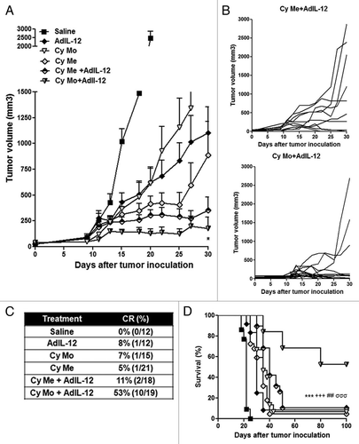
Table 1. Analysis of in vivo antitumor synergy by the fractional product method.
Figure 2. (A) Quantification of IL-10. Serum IL-10 was measured in CT26 tumor-bearing mice treated with saline, metronomic cyclophosphamide (Cy Me, 25 mg/kg i.p., 3 times/week, from day 8), or single low-dose cyclophosphamide (Cy Mo, 50 mg/kg i.p., day 8). Data are expressed as mean (bars = SEM) serum concentration.***p < 0.001: Cy Mo vs. saline ; ### p < 0.001 Cy Me vs. saline. (B) Percentage of CD4+FOXP3+ cells (Tregs) in peripheral blood. ** p < 0.01: Cy Mo + AdIL-12 vs. saline; # p < 0.05: Cy Me + AdIL-12 vs. saline; (C) or spleen ** p < 0.01: Cy Mo + AdIL-12 vs. saline; ## p < 0.01: Cy Me + AdIL-12 vs. saline. (D) Percentage of splenic CD11b+Gr1+ cells at days 7 and (E) at 15 post treatment). Day 7: *** p < 0.001 Cy Mo + AdIL-12 vs. saline; ### Cy Me+AdIL-12 vs. saline. Day 15: *** p < 0.001 Cy Mo + AdIL-12 vs. saline; # p < 0.05 Cy Mo + AdIL-12 vs. Cy Me + AdIL-12. One-way ANOVA and Bonferroni post-test were used to compare differences among groups.
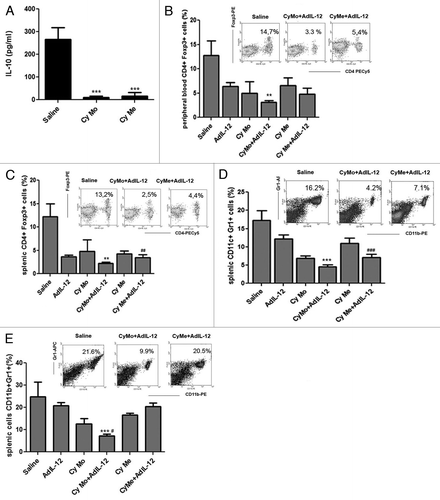
Figure 3. (A) Single low-dose cyclophosphamide enhances allogeneic splenocyte proliferation. Splenocytes from C57BL/6 mice were stimulated in vitro with DCs pulsed with tumor extracts from CT26-bearing mice treated with single low-dose cyclophosphamide (Cy Mo) or saline during 5 d. Cell proliferation was evaluated by 3H-thymidine incorporation assay. ** p < 0.01: DCCyMo vs. DCsaline. (B) IFNγ quantification in supernatants from cell proliferation assay. ** p < 0.01: DCCyMo vs. DCsaline. (C) Percentage of MHC-II+CD80+ cells in CD11c+ gate from pulsed DCCyMo or DCsaline. * p < 0.05: DCCyMo vs. DCsaline. (D) IL-12 production from pulsed DCCyMo or DCsaline. * p < 0.05: DCCyMo vs. DCsaline (E) Allogeneic splenocytes proliferation induced by both treatment with single low-dose or metronomic cyclophosphamide. Splenocytes from C57BL/6 mice were stimulated in vitro with DCs pulsed with tumor extracts obtained from CT26-bearing mice treated with Cy Mo, metronomic cyclophosphamide (Cy Me) or saline during 5 d. Cell proliferation was evaluated as described above. * p < 0.05: DCCyMo vs. DCCyMe. Kruskal–Wallis and Dunn’s test were used to compare differences among groups.
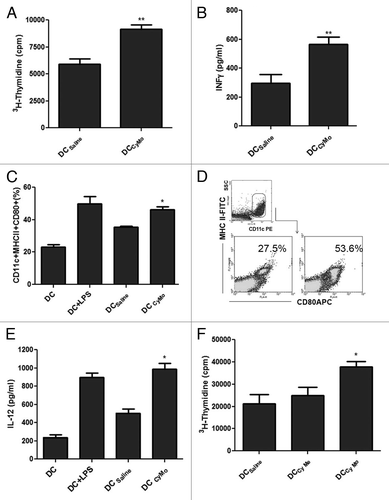
Figure 4. (A) Single low-dose cyclophosphamide is superior to metronomic cyclophosphamide in inducing specific CTL activity. Splenocytes from different experimental groups were stimulated in vitro with mitomycin C–treated CT26 cells for 5 d. Specificity of CTL cytotoxicity was evaluated using BNL cells. The percentage of specific cytotoxicity was calculated according to the following formula: [(abs 492 nm experimental - abs 492 nm background)/(abs 492 nm maximum - abs 492 nm background)] × 100. Data represent the mean of cuatriplicate cultures. * p < 0.05: single low-dose cyclophosphamide (Cy Mo) + AdIL-12 vs. Cy Mo, AdIL-12 or metronomic cyclophosphamide (Cy Me) + AdIL-12. One way ANOVA and Tukey’s multiple comparison post-test was used to compare differences among groups. (B) Tumor-infiltrating T cells labeled with anti-CD3 antibodies were analyzed by immunohistochemistry (200X). (C) Percentage of intratumoral CD4+ and (D) CD8+ cells, as analyzed by flow cytometry. *p < 0.05: Cy Mo + AdIL-12 vs. Cy Me + AdIL-12.
![Figure 4. (A) Single low-dose cyclophosphamide is superior to metronomic cyclophosphamide in inducing specific CTL activity. Splenocytes from different experimental groups were stimulated in vitro with mitomycin C–treated CT26 cells for 5 d. Specificity of CTL cytotoxicity was evaluated using BNL cells. The percentage of specific cytotoxicity was calculated according to the following formula: [(abs 492 nm experimental - abs 492 nm background)/(abs 492 nm maximum - abs 492 nm background)] × 100. Data represent the mean of cuatriplicate cultures. * p < 0.05: single low-dose cyclophosphamide (Cy Mo) + AdIL-12 vs. Cy Mo, AdIL-12 or metronomic cyclophosphamide (Cy Me) + AdIL-12. One way ANOVA and Tukey’s multiple comparison post-test was used to compare differences among groups. (B) Tumor-infiltrating T cells labeled with anti-CD3 antibodies were analyzed by immunohistochemistry (200X). (C) Percentage of intratumoral CD4+ and (D) CD8+ cells, as analyzed by flow cytometry. *p < 0.05: Cy Mo + AdIL-12 vs. Cy Me + AdIL-12.](/cms/asset/30c5bffe-195b-4cbc-bc68-46b088d27899/koni_a_10920684_f0004.gif)