1,880
Views
55
CrossRef citations to date
0
Altmetric
Original Research

Expression of MHC Class I on breast cancer cells correlates inversely with HER2 expression

, , , , , , , , , , , , , , & show all
Pages 1104-1110 | Received 01 Mar 2012, Accepted 08 Jun 2012, Published online: 01 Oct 2012

Figures & data

Figure 1. Representative immunostaining of HER2, MHC class I and PTEN. (A) HER2 staining (0, 1+, 2+, 3+). (B) MHC class I staining (weak and strong). (C) PTEN staining (0, 1+, 2+, 3+). Original magnification: 200x.
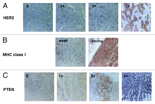
Figure 1. Representative immunostaining of HER2, MHC class I and PTEN. (A) HER2 staining (0, 1+, 2+, 3+). (B) MHC class I staining (weak and strong). (C) PTEN staining (0, 1+, 2+, 3+). Original magnification: 200x.

Table 1. Clinical features of the patients (n = 70)

Figure 2. Representative MHC class I immunostaining of HercepTest 0 (A) and 3+ (B) cases. Serial sections were stained with antibodies specific for HER2 and MHC class I. (A) MHC class I staining was strong in HercepTest 0 case. (B) MHC class I staining was weak in HercepTest 3+ case. Original magnification: 200x.
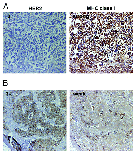
Figure 2. Representative MHC class I immunostaining of HercepTest 0 (A) and 3+ (B) cases. Serial sections were stained with antibodies specific for HER2 and MHC class I. (A) MHC class I staining was strong in HercepTest 0 case. (B) MHC class I staining was weak in HercepTest 3+ case. Original magnification: 200x.

Table 2. Summarized immunohistochemical staining data (n = 70)

Figure 3. HER2 downregulation by siRNA upregulates MHC class I expression on breast cancer cells. MDA-MB-231 cells were transfected with a non-targeting siRNA (siCont) or with a siRNA for the downregulation of HER2 (siHER2). HER2 expression was downregulated in siHER2 transfectants, as assessed by flow cytometry (A) and immunoblotting (B). (C) MHC class I expression was increased in cells transfected with siHER2, as assessed by cytofluometry upon staining with two different antibodies specific for MHC class I molecules, clone W6/32 and clone G46–2.6. Representative results from n = 5 independent experiments are shown.
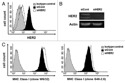
Figure 3. HER2 downregulation by siRNA upregulates MHC class I expression on breast cancer cells. MDA-MB-231 cells were transfected with a non-targeting siRNA (siCont) or with a siRNA for the downregulation of HER2 (siHER2). HER2 expression was downregulated in siHER2 transfectants, as assessed by flow cytometry (A) and immunoblotting (B). (C) MHC class I expression was increased in cells transfected with siHER2, as assessed by cytofluometry upon staining with two different antibodies specific for MHC class I molecules, clone W6/32 and clone G46–2.6. Representative results from n = 5 independent experiments are shown.

Table 3. HER2-siRNA effect on MHC class I expression

Figure 4. MAPK inhibiton upregulates MHC class I expression on breast cancer cells. MDA-MB-231, BT549 and SK-BR-3 were treated with the ERK inhibitor PD98059. Phospho-p44MAPK and phospho-p42MAPK levels were reduced in a dose-dependent manner, as assessed by immunoblotting. Conversely, MHC class I expression was increased upon exposure to PD98059 in a dose-dependent manner, as assessed by cytofluometry upon staining with two different antibodies specific for MHC class I molecules, clone W6/32 and clone G46–2.6. Non-viable (7-AAD+) cells were excluded from the analysis. Representative results from n = 4 independent experiments are shown. MFI, mean fluorescence intensity.
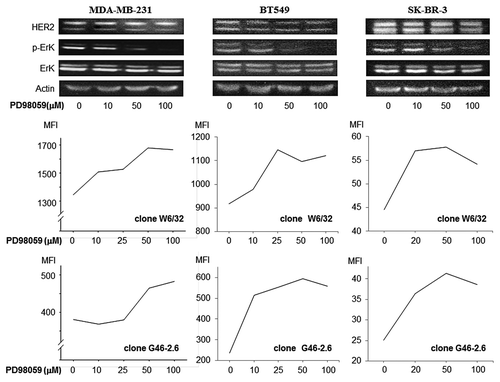
Figure 4. MAPK inhibiton upregulates MHC class I expression on breast cancer cells. MDA-MB-231, BT549 and SK-BR-3 were treated with the ERK inhibitor PD98059. Phospho-p44MAPK and phospho-p42MAPK levels were reduced in a dose-dependent manner, as assessed by immunoblotting. Conversely, MHC class I expression was increased upon exposure to PD98059 in a dose-dependent manner, as assessed by cytofluometry upon staining with two different antibodies specific for MHC class I molecules, clone W6/32 and clone G46–2.6. Non-viable (7-AAD+) cells were excluded from the analysis. Representative results from n = 4 independent experiments are shown. MFI, mean fluorescence intensity.