Figures & data
Figure 1. Gr-1+CD11b+ cells expand during cancer progression in nude mice as in immunocompetent balb/c mice. Balb/c and nude mice were injected in the 4th mammary fat pad with 4T1 and MDA-MB-231 breast cancer cells respectively or PBS as a control. After 4 weeks of tumor growth, mice were sacrificed. Spleen and BM cells were isolated and Gr-1+CD11b+ cells expansion examined in tumor bearing mice. (A) Spleen and bone marrow of control and tumor nude mice were analyzed by flow cytometry. (B) Quantitative analysis of the expansion of Gr-1+CD11b+ cells in spleens and bone marrow of balb/c and nude mice 4 weeks after mammary fat pad inoculation of 4T1 or MDA-MB-231 breast cancer cells respectively. In both strands, control mice were injected with PBS. Results are presented as the mean ± SEM (5 mice per group)
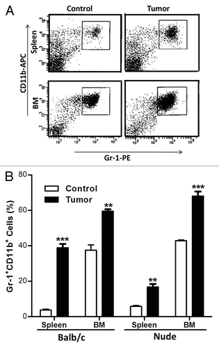
Figure 2. Tumor-induced Gr-1+CD11b+ cells promote tumor cells growth and impair bone mass. One × 106 MDA-MB-231 and 1 × 10⁵ Gr-1+CD11b+ cells isolated from the spleen of control or tumor mice were co-injected in the 4th mammary fat pad of nude mice. Tumor growth was monitored by caliper measurement every 2–3 d (A). At day 24, mice were sacrificed, tumors removed, measured (B) and weighed (C). Results were presented as the mean ± SEM (10 mice per group). (D) Bone mass was assessed by microCT analysis of right femurs from both groups. Data are presented as the mean ± SEM (8 to 10 mice per group).
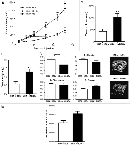
Figure 3. Tumor-induced Gr-1+CD11b+ cells promote bone metastasis in a model of breast cancer metastasis to bone. One × 105 MDA-MB-231 were inoculated into the left cardiac ventricle in nude mice and 1 × 10⁵ Gr-1+CD11b+ cells isolated from the BM of control or tumor mice were injected in the left tibia of the same mice. The right tibia was injected with PBS as a control. (A) Quantitative analysis of GFP fluorescence detected in mice long bones. (B) Quantitative analysis of osteolytic lesions observed in long bones after radiographs (C) Representative pictures of TRAP staining on bone sections from co-injected mice (left panel) after sacrifice at week 4, and quantitative analysis of the ratio bone surface/osteoclast number (right panel). (D) Osteolytic lesions area were quantified separately in right and left hindlimbs for each mouse.
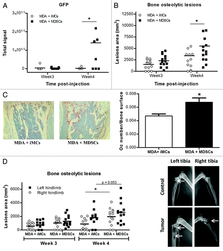
Figure 4. Tumor-induced MDSCs lose their ability to decrease bone mass in the absence of tumor cells. 1 × 10⁵ Gr-1+CD11b+ cells isolated from the BM of control or tumor mice were injected in intratibial in the left tibia. Right tibiaes of the same mice were injected with PBS as a control. Bone parameters were assessed by μCT
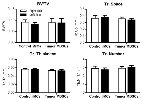
Figure 5. Gr-1+CD11b+ cells expand in a model of breast cancer metastasis to bone and acquire a phenotype that can promote osteolysis. (A) 105 MDA-MB-231 or PBS were injected in IC in nude mice. After 2 and 4 weeks, mice were sacrificed and the Gr-1+CD11b+ cell population in spleen and BM analyzed by flow cytometry. The Gr-1lowCD11b+ population was also analyzed at week4 in BM. (B) Gr-1+CD11b+ were isolated from the BM of control or tumor mice using MACS magnetic microbeads cell sorting and their purity checked. (C-D) Gr-1+CD11b+ cells sorted from control or tumor mice were plated in 6 wells plates and co-cultured with MDA-MB-231 plated in transwells. After 48h, RNA was extracted from both cell types; PTHrP and Gli2 expression in MDA-MB-231 cells (C) and TGF-β expression in Gr-1+CD11b+ cells (D) were analyzed by qRT-PCR.
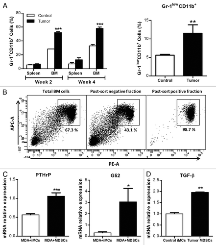
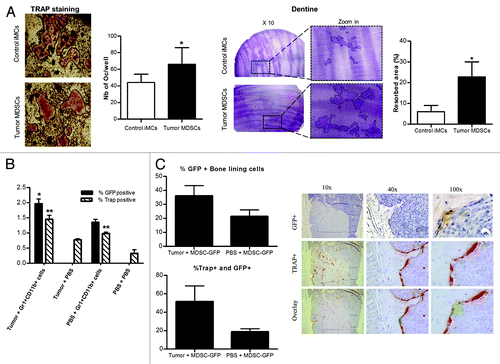
Figure 6. Gr1+CD11b+ cells differentiate into osteoclasts in vitro and in vivo. (A) Sorted MDSCs were plated in 48 well plates with dentine discs and cultured in vitro with 25ng/ml of M-CSF and 50ng/ml of RANKL for about 14 d. Wells and dentine discs were then stained and the number of osteoclasts and the resorption area (circled on the zoom panel) quantified. (B) 105 Gr-1+CD11b+ cells isolated from the BM of GFP mice were injected into the left tibia of mice. Mice were co-injected with 106 MDA-MB-231 breast cancer cells or PBS in intracardiac. Four weeks after injections, mice were sacrificed and bones collected for immunostaining with a GFP antibody and for TRAP staining (4 to 8 mice per group). The percentage of total BM positive cells for each staining is represented on the graph (C) Percentage of bone lining cells and TRAP positive cells among GFP+ cells from (B) (left panel). A representative picture of a GFP+ osteoclast is presented on the right panel.