Figures & data
Figure 1. Sympathetic stellate ganglion; most cardiac-projecting neurons have their origins in this ganglion. Developmental profile with immunohistochemistry for the noradrenergic marker tyrosine hydroxylase (TH; green) in sheep. Sheep sympathetic neurons express TH by E100 with more prominent expression by E129; staining is localized in neuronal cytoplasm (white arrowheads) and within nerve bundles and fibers (gray arrowheads) at all ages examined (Jonker S., Louey S., Moses M., Macek A., Giraud G., Thornburg K., Hasan W.; unpublished data). Scale bar in C is 200 υm.
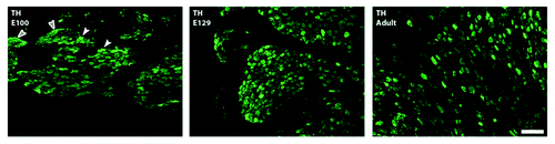
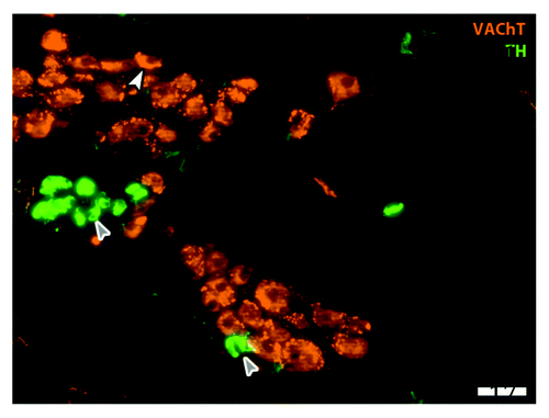
Figure 2. Parasympathetic cardiac ganglion. Immunohistochemical staining in an adult rat cardiac ganglion for cholinergic marker vesicular acetylcholine transporter (VAChT; red) and catecholaminergic marker tyrosine hydroxylase (TH; green). Neurons stain in the soma for VAChT (white arrowhead) and also receive input from cholinergic pre-ganglionic terminals (small varicosities around cell bodies). Small intensely fluorescent (SIF; gray arrows) cells are catecholaminergic and believed to function as inter-neurons; here SIF cells make contact with each other and with a cholinergic neuron (Moses M., Macek A. Hasan W.; unpublished data). Scale bar is 50 υm.