1,136
Views
12
CrossRef citations to date
0
Altmetric
Review

Small non-coding RNAs in streptomycetes

, &
Pages 464-469 | Received 27 Jan 2014, Accepted 17 Feb 2014, Published online: 06 Mar 2014

Figures & data

Figure 1. Complex life cycle of streptomycetes. (A) Morphological diversity of different Streptomyces strains. (B) The life cycle of S. coelicolor. Monogenomic spores of S. coelicolor germinate and grow into the soil. When nutrition becomes limited the vegetative mycelia digest themselves to serve as substrate for aerial mycelia growing out of the soil. Those aerial hyphae differentiate into spores again through insertion of septa.
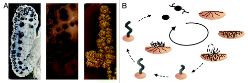
Figure 1. Complex life cycle of streptomycetes. (A) Morphological diversity of different Streptomyces strains. (B) The life cycle of S. coelicolor. Monogenomic spores of S. coelicolor germinate and grow into the soil. When nutrition becomes limited the vegetative mycelia digest themselves to serve as substrate for aerial mycelia growing out of the soil. Those aerial hyphae differentiate into spores again through insertion of septa.

Figure 2. Agarase DagA controlled by scr5239. (A) Agarase assay of S. coelicolor wild-type, scr5239 deletion, and overexpression strain. Agarase expression is visualized as a pale halo around the colony. (B) Electrophoretic mobility shift of radiolabelled scr5239 (open circle) incubated with an increasing amount of a 100 nt mRNA fragment containing the target site of dagA. Complex formation (closed circle) was visualized by gelelectrophoresis. (C) Secondary structure of scr5239 alone (top) and in complex with the mRNA target site (bottom). The structure was calculated using RNAfoldCitation29 and confirmed by structural probing. Stems (pedesand = P) are numbered, the 5′ and 3′ end of each RNA is indicated.
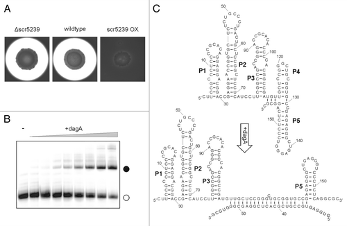
Figure 2. Agarase DagA controlled by scr5239. (A) Agarase assay of S. coelicolor wild-type, scr5239 deletion, and overexpression strain. Agarase expression is visualized as a pale halo around the colony. (B) Electrophoretic mobility shift of radiolabelled scr5239 (open circle) incubated with an increasing amount of a 100 nt mRNA fragment containing the target site of dagA. Complex formation (closed circle) was visualized by gelelectrophoresis. (C) Secondary structure of scr5239 alone (top) and in complex with the mRNA target site (bottom). The structure was calculated using RNAfoldCitation29 and confirmed by structural probing. Stems (pedesand = P) are numbered, the 5′ and 3′ end of each RNA is indicated.

Reprints and Corporate Permissions

Please note: Selecting permissions does not provide access to the full text of the article, please see our help page How do I view content?

To request a reprint or corporate permissions for this article, please click on the relevant link below:

Academic Permissions

Please note: Selecting permissions does not provide access to the full text of the article, please see our help page How do I view content?

Obtain permissions instantly via Rightslink by clicking on the button below:

If you are unable to obtain permissions via Rightslink, please complete and submit this Permissions form. For more information, please visit our Permissions help page.