Figures & data
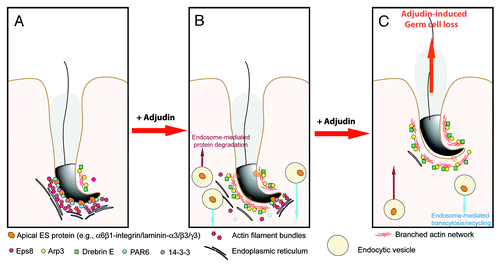
Figure 1. A summary of effects of adjudin that impede spermatogenesis based on studies in the rat. This figure summarizes recent findings in the field regarding the effects of adjudin on different proteins, protein complexes, or cellular structures in the seminiferous epithelium of rat testes. While testin and actin are the molecular targets of adjudin,Citation23 it is not known at present if testin is also involved in the adjudin-mediated effects on the homeostasis of proteases/protease inhibitors and the actin-based cytoskeletal at the apical ES. This figure was prepared based on recent reviews.Citation12,Citation24-Citation29 MMP2, matrix metalloproteinase 2; α2-MG, α2-macroglobulin; TIMPs, tissue metalloproteinases.
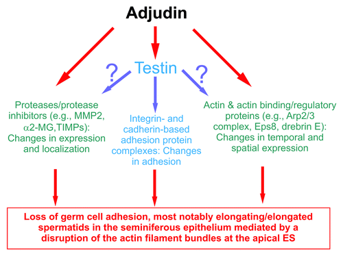
Figure 2. Adjudin disrupts the restricted temporal and spatial expression of Eps8 and Arp3 in the rat testis. Dual-labeled immunofluorescence analysis was used to assess changes in the adjudin-mediated disruption of the highly restricted spatial and temporal expression of Eps8 (green) and F-actin (red) shown in (A) vs. Arp3 (green) and F-actin shown in (B) in the seminiferous epithelium of rat testes as earlier described.Citation32,Citation33 In both (A) and (B), the control cross-sections illustrate a stage VII tubule. By 12h (hour) following adjudin treatment, a considerable loss of Eps8 was detected at the apical ES, and by 1D (day), Eps8 was virtually not detected, and this loss of Eps8, an actin bundling protein at the apical ES, was found to associate with a truncation of F-actin due to the loss of the organized actin filament bundles at the apical ES (see e-f and g-i vs. a-c in A). For Arp3, it displayed a distinctive different pattern vs. Eps8 following adjudin treatment. While both Eps8 and Arp3 were prominently expressed at the apical ES at stage VII in control rats (see a-c in B vs. a-c in A), however, Arp3 was highly restricted to the concave side of the spermatid head where endocytic vesicle-mediated protein trafficking is known to occur.Citation40 By 12h following adjudin treatment, Arp3 became mis-localized and this mis-localization was more obvious by 1D after adjudin treatment. For instance, by 1D, Arp3 was found to become truncated (see “white” arrowheads in the enlarged inset in B:g). Additionally, Arp3 was also found to be mis-localized, surrounding other parts of the spermatid head, instead of restricted to the concave side of the spermatid head (see “yellow” arrowheads in the enlarged inset in B:g). Thus, the actin filament bundles at the apical ES were transformed to a branched network because Arp3 induces actin nucleation/branching. The combined changes of these two actin regulatory proteins induced by adjudin thus led to the degeneration of the actin filament bundles at the apical ES. These changes coupled with changes in drebrin E, PAR6 and 14–3-3 as described in the main text destabilizes the apical ES, rendering a loss of adhesion at the apical ES which led to spermatid loss from the epithelium. Bar in A:a or B:a = 40 μm, which applies to b-i in A or B; bar in inset in B:a = 20 μm, which applies to insets in b-c and g-i.
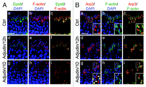
Figure 3. A hypothetical model that summarizes the mechanism by which adjudin induces spermatid loss from the seminiferous epithelium of adult rat testes. In a normal rat testis (A), apical ES adhesion is maintained by the apical ES adhesion protein complexes (e.g., α6β1-integrin-laminin-α3β3γ3, N-cadherin-β-catenin, nectin-2/3-afadin) using the highly organized actin filament bundles for attachment. The actin filament bundles at the apical ES are maintained by the Eps8, PAR6 and 14–3-3. However, following exposure of rats to adjudin, this drug disrupts the highly restricted temporal and spatial expression of Eps8, Arp3, drebrin E, PAR6 and 14–3-3 in the seminiferous epithelium as detailed in the text. In short, Eps8 considerably diminishes at the apical ES with a concomitant mis-localization and truncation of Arp3, the actin filament bundles thus become disrupted and replaced with actin branched network. At the same time, PAR6 and 14–3-3 also become diminished, accelerating endocytic vesicle-mediated internalization of integral membrane proteins (e.g, integrins, cadherins, nectins), thereby destabilizing the apical ES (B), which eventually leads to premature loss of spermatid from the epithelium, mimicking spermiation as shown in (C).