Figures & data
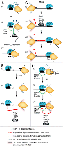
Figure 1 tRNA genes and the transcriptional machinery. (A) Alignment of three of ten wild-type budding yeast genes in the tL(CAA) tRNA gene family (also called the Leu3 family). The leucine anticodon is boxed and the intron is yellow. The genes are identical in their coding sequences. Also shown is the sequence of SUP53, which results from an A to T mutation (orange) in the anticodon of the wild-type tL(CAA)C gene. SUP53 is a leucine-inserting amber suppressor. (B) Sequence of tL(CAA)C showing the locations of the A and B boxes (blue), which are bound by TFIIIC. Transcription initiates at the arrow. The terminator is a run of five T residues (purple) just downstream of the coding region. (C) The core components of the tRNA gene transcriptional apparatus in budding yeast and their human counterparts (tabulated).Citation4–Citation6
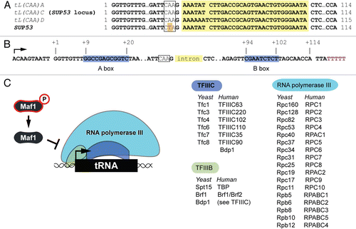
Figure 2 Replication interference by tRNA genes. The straight arrow shows the direction of transcription. The curved arrows show the direction of polymerase movement during nucleic acid synthesis. Curved T icons represent inhibitory effects on replication.
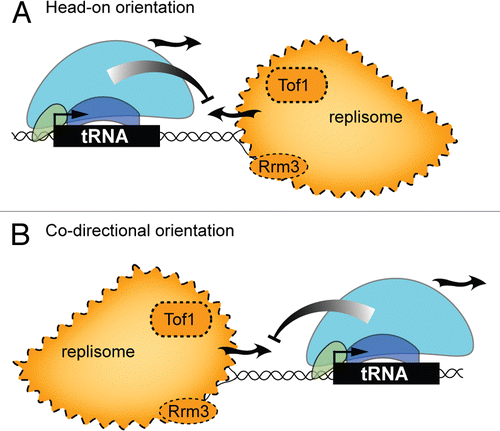
Figure 3 Control of tRNA gene transcription in yeast by replication stress checkpoint signaling molecules. In this model the checkpoint proteins are organized according to dependencies established in the literature. Dashed lines represent signaling steps that are yet to be fully described. ssDNA, single strand DNA.
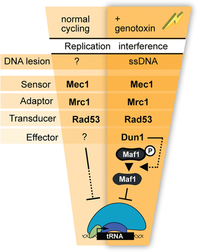
Figure 4 Mechanism of initiation and spatial range of checkpoint signaling to the tRNA genes. The models depict events that occur during normal cycling (A), and under conditions of replication stress induced by HU and MMS (B–D). The scenarios outlined include cis-signaling where a fork is paused at a tRNA gene (A-i and D), trans-signaling from a fork paused at a tRNA gene to another tRNA gene (A-ii), and trans-signaling from a stalled fork to a distal tRNA gene (B and C). The curved arrow represents the direction of fork movement. The PIC on tRNA genes is labeled ”III-B-C.”