1,531
Views
59
CrossRef citations to date
0
Altmetric
Review

Skeletal myogenesis and Myf5 activation

&
Pages 109-114 | Received 15 Mar 2011, Accepted 13 Apr 2011, Published online: 01 May 2011

Figures & data

Figure 1 Myogenesis in the somite is regulated by signaling molecules from neighboring tissues and factors expressed by MPCs. Epaxial myogenesis is positively regulated by Wnt from neural tube and Shh from floor plate and notochord. Wnt signals from dorsal ectoderm induce myogenesis in hypaxial myotome, whereas BMP from lateral mesoderm inhibits. Pax 3/7, Six 1/4, Eya 1/2 expressed by MPCs positively regulate myogenesis.
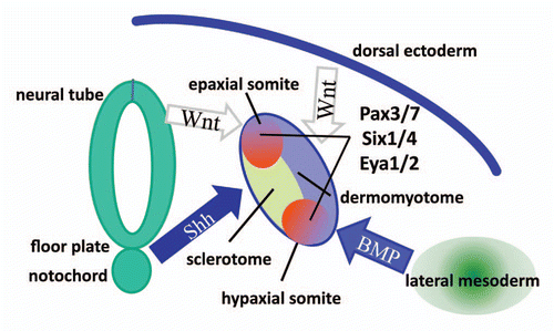
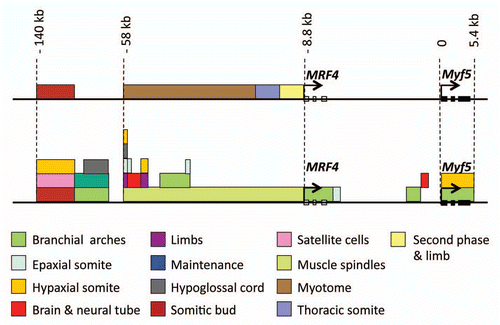
Figure 1 Myogenesis in the somite is regulated by signaling molecules from neighboring tissues and factors expressed by MPCs. Epaxial myogenesis is positively regulated by Wnt from neural tube and Shh from floor plate and notochord. Wnt signals from dorsal ectoderm induce myogenesis in hypaxial myotome, whereas BMP from lateral mesoderm inhibits. Pax 3/7, Six 1/4, Eya 1/2 expressed by MPCs positively regulate myogenesis.

Figure 2 Myf5 and MRF4 genes are both located on chromosome 10, about 8.8 kb apart. Elements regulating MRF4 located upstream of MRF4 start site are shown on the top part. Myf5 enhancers spanning 140 kb upstream of Myf5 start site and the intragenic region of Myf5 are shown on the bottom part.

Figure 2 Myf5 and MRF4 genes are both located on chromosome 10, about 8.8 kb apart. Elements regulating MRF4 located upstream of MRF4 start site are shown on the top part. Myf5 enhancers spanning 140 kb upstream of Myf5 start site and the intragenic region of Myf5 are shown on the bottom part.