551
Views
10
CrossRef citations to date
0
Altmetric
Editor's Corner

Fungal x host interactions in Chromoblastomycosis

What we have learned from animal models and what is yet to be solved

Pages 3-5 | Received 10 Aug 2009, Accepted 24 Sep 2009, Published online: 28 Feb 2010

Figures & data

Figure 1 Sclerotic cells can be isolated from human lesions and from plants. Typical round, thick-walled, brown color, septated (arrows) sclerotic cells after direct mycological examination from human lesion (A) or from Mimosa pudica thorns (B). scale bars: 10 µm.
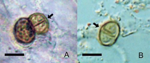
Figure 1 Sclerotic cells can be isolated from human lesions and from plants. Typical round, thick-walled, brown color, septated (arrows) sclerotic cells after direct mycological examination from human lesion (A) or from Mimosa pudica thorns (B). scale bars: 10 µm.

Figure 2 In vitro-generated sclerotic cells can stimulate the formation of Langhans giant cells in vitro. Langhans giant cells are observed 24 hours after interaction with balb/c mice peritoneal macrophages (A). Morphologically similar giant cells are present in the histopathology of lesional skin (B). Scale bars: 50 µm.
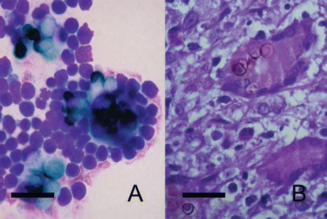
Figure 2 In vitro-generated sclerotic cells can stimulate the formation of Langhans giant cells in vitro. Langhans giant cells are observed 24 hours after interaction with balb/c mice peritoneal macrophages (A). Morphologically similar giant cells are present in the histopathology of lesional skin (B). Scale bars: 50 µm.