Figures & data
Figure 1 Common histopathological findings of footpad tissue from mice stimulated at two sites; H&E. Focal lesions that were granuloma-like (A, 40x) and rich in fungal cells (B, 400x) observed 30 days post-infection. (C) After 60 days of infection, lesions presented low numbers of sclerotic-like cells (arrows; 200x and insert 1000x), regions rich in histiocytes*H (clear cytoplasms due to cell-cell junctions) and organized focal lesions surrounded by fibroblasts*F (cells with elongated nucleus). (D) 90 days post-infection, high numbers of polymorphonuclear cells(*) were verified in focal lesions without the presence of fungal cells.
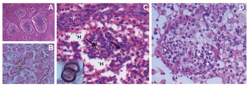
Figure 2 Metastasis of sclerotic-like cells in animals stimulated at two sites. (A) Mouse infected in the footpad with F. pedrosoi cells; the animal received a simultaneous immunization i.p. with heat-killed fungal cells. (B) Common morphology of sclerotic-like cells that migrated to other sites, 1000x.
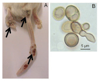
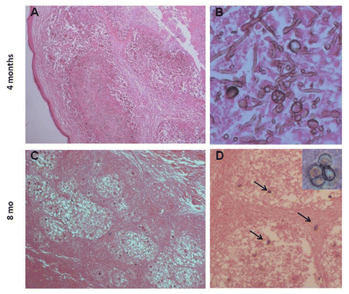
Figure 3 Histopathological characteristics of focal lesions in Xid mice 4 and 8 months post-costimulation; H&E. Larger and irregular lesions of the footpad (A, 100x) with high numbers of thin, small hyphae, conidia and conidiogenous cells (B, 400x) four months post-infection. Small and well-organized granuloma-like lesions (C, 100x) with a small number of sclerotic-like cells (D, 200x and insert 1000x) were observed after eight months.
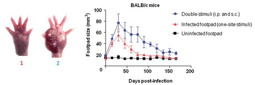
Graph 1 Co-stimulation of the footpad and the peritoneum caused an increase in footpad swelling as well as a prolonged infection in BALB/c mice. The footpads of mice were measured every 15 days for 165 days after inoculation with F. pedrosoi. Animals infected s.c. in the footpad with viable F. pedrosoi cells (triangles) generally presented an infection peak after 30 days and were healed after 60 days post-infection, whereas the lesions of mice co-stimulated by immunization i.p. with nonviable cells and a s.c. footpad infection with fungus had strong swelling and a slower healing rate (circles). The control group of animals was treated with 50 µL of PBS (uninfected footpad, squares). Data are shown with mean and SE values; Student's t test (p < 0.001). Images of the footpads of mice stimulated at a single site (1) and at two sites (2) are shown at 30 days post-infection (left of the graph).
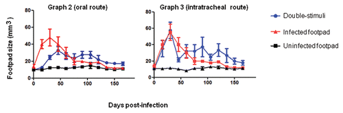
Graphs 2 and 3 Kinetics of footpad lesions in BALB/c mice infected with viable F. pedrosoi cells that received oral (graph 2) or pulmonary (graph 3) mucosal stimuli with nonviable fungal cells. Data are shown as the mean +/- SE. The Student's t test was used to verify the significance between co-stimulation and footpad infection (one site) data; p > 0.05.
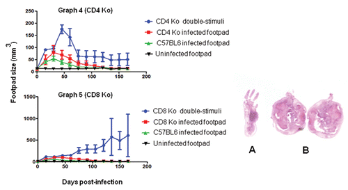
Graphs 4 and 5 CD4 KO and CD8 KO mice that were footpad infected with viable F. pedrosoi cells and co-stimulated i.p. with heat-killed fungal cells (co-stimulated). In the early periods post-infection, increased swelling was observed in the footpads of the two animal groups. Animals in the CD4 KO group had a decrease in inflammation after 60 days (). CD8 KO mice did not heal and showed a strong progressive enlargement of the footpad (). Data are shown with mean and SE values; an ANOVA test was used for and ; p < 0.05. In the right panel are pictures of histopathological sections from the footpad of a one-site infected CD8 KO mouse at 30 days (A) and enlarged footpads after 150 days from CD8 KO mice that were co-stimulated (B).
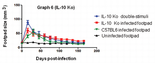
Graph 6 Co-stimulated IL-10 KO mice presented with progressive inflammation of the footpads early after infection and healed after 60 days; this was similar in all animal groups. A one-way ANOVA was performed for the IL-10 group; p > 0.05.
Graphs 7 and 8 Deficiency in B1 B cells causes an increase of host susceptibility to F. pedrosoi infection in co-stimulated mice. Co-stimulated Xid were chronically infected (). Immune control of the fungus in irradiated animals generally occurs after 90 days post-infection (). Data are shown with mean and SE values. An ANOVA was used for analyses of significance for Xid mice (p < 0.05), and the Student's t test was used for the irradiated mice as well as the BALB/c footpad infection data (p = 0.0304).
