Figures & data
Figure 1 Molecular Koch's postulates dissection of toxin contributions to the virulence of type C isolate CN3685 in rabbit ileal loops. In this model, wild-type CN3685 causes necrotizing enteritis, with an accumulation of bloody fluid inside the ileal loops. Similar pathology was observed for isogenic mutants unable to produce perfringolysin O (pfoA), α-toxin (plc), or lacking both perfringolysin O and α-toxin (plc/pfoA). In contrast, an isogenic mutant (cpb) unable to produce β-toxin was completely attenuated for virulence; this virulence was restored by complementation (not shown in ). also shows that purified β-toxin alone can reproduce the pathology caused by wild-type CN3685 infection. Modified and used with permission from Sayeed et al.Citation3
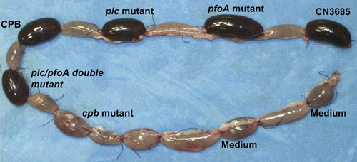
Figure 2 The presence of host cells affects the kinetics of β-toxin accumulation in supernatants of type C isolate JGS1495. Equal number of JGS1495 cells were inoculated into a tissue culture dish containing bacterial culture media [either fluid thioglycollate broth (FTG) or tryptic soy broth-glucoseyeast extract (TGY)] tissue culture medium [minimal essential medium (MEM)], or MEM containing Caco-2 enterocyte-like cells. After 3 h incubation at 37°C, each culture was harvested and the culture supernatant was subjected to western blotting using an anti-β-toxin monoclonal antibody. Used with permission from Vidal et al.Citation11
![Figure 2 The presence of host cells affects the kinetics of β-toxin accumulation in supernatants of type C isolate JGS1495. Equal number of JGS1495 cells were inoculated into a tissue culture dish containing bacterial culture media [either fluid thioglycollate broth (FTG) or tryptic soy broth-glucoseyeast extract (TGY)] tissue culture medium [minimal essential medium (MEM)], or MEM containing Caco-2 enterocyte-like cells. After 3 h incubation at 37°C, each culture was harvested and the culture supernatant was subjected to western blotting using an anti-β-toxin monoclonal antibody. Used with permission from Vidal et al.Citation11](/cms/asset/e6cd6588-4d34-4375-b540-96044c609fbb/kvir_a_10910679_f0002.gif)
Table 1 Toxintyping classification of C. perfringens isolates