Figures & data
Figure 1 Eukaryotic SNAREs and viral coiled-coil proteins trigger membrane fusion. (A) During a fusion event in eukaryotic cells, the t-SNAREs present on the target membrane interact with the v-SNAREs present on the vesicle. This interaction brings both membranes into a close apposition and leads to membrane fusion. (B) During a viral infection, enveloped viruses enter their host cells through fusion with the host membrane (plasma membrane or endosome membrane). For example, HIV envelope protein gp41 utilizes three α-helical domains, which collapse into a trimer of hairpins to facilitate fusion between the host plasma membrane and the viral envelope thereby providing its viral contents access to the host cytosol.
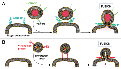
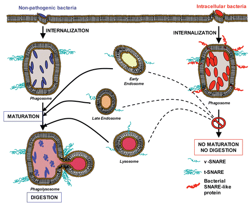
Figure 2 Bacterial coiled-coil proteins inhibit membrane fusion (working model). Left: Non-pathogenic bacteria (blue) are internalized. Normal phagosome maturation is initiated and results in lysosomal fusion (formation of phagolysosomes) and destruction of the phagosomal content. Right: Intracellular bacteria (red) are internalized and express their own proteins on the surface of the phagosome (red coiled-coil proteins), blocking its maturation.