3,917
Views
110
CrossRef citations to date
0
Altmetric
Review

Targeting of the central nervous system by Listeria monocytogenes

&
Pages 213-221 | Published online: 01 Mar 2012

Figures & data

Figure 1. Blood-CSF and blood-brain barriers. From top to bottom: (A) Capillaries from the meningeal compartment, which represent part of the blood-CSF barrier. Dural endothelial cells are fenestrated, while endothelial cells from the subarachnoid space are joined by tight junctions. (B) Blood-brain barrier is made of endothelial cells joined by tight junctions. They are in close contact with astrocyte’s “feet” (astrocyte’s cell projection), which enable the differentiation of brain endothelial cell junctions. (C) Epithelial cells from the choroid plexus produce the cerebrospinal fluid (CSF) and isolate the fenestrated endothelium from the CSF, hence forming together with the meningeal endothelium the blood-CSF barrier. (Adapted from ref. Citation64.)
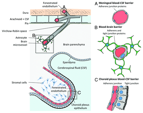
Figure 1. Blood-CSF and blood-brain barriers. From top to bottom: (A) Capillaries from the meningeal compartment, which represent part of the blood-CSF barrier. Dural endothelial cells are fenestrated, while endothelial cells from the subarachnoid space are joined by tight junctions. (B) Blood-brain barrier is made of endothelial cells joined by tight junctions. They are in close contact with astrocyte’s “feet” (astrocyte’s cell projection), which enable the differentiation of brain endothelial cell junctions. (C) Epithelial cells from the choroid plexus produce the cerebrospinal fluid (CSF) and isolate the fenestrated endothelium from the CSF, hence forming together with the meningeal endothelium the blood-CSF barrier. (Adapted from ref. Citation64.)

Figure 2. Cranial nerves in human. A frame surrounds the nerves that emerge from the regions most frequently infected by Lm in the brainstem. Adapted from Patrick Lynch; Creative Commons Attribution 2.5 License 2006; www.patricklynch.net.
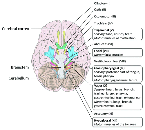
Figure 2. Cranial nerves in human. A frame surrounds the nerves that emerge from the regions most frequently infected by Lm in the brainstem. Adapted from Patrick Lynch; Creative Commons Attribution 2.5 License 2006; www.patricklynch.net.

Figure 3. Potential mechanisms by which Lm could cross the blood-brain barrier. (A) Extracellular Lm, either free in the blood and/or associated to circulating cells, may recognize receptors at the surface of the barriers (as InlA, InlB or Vip) and cross them. (B) Trojan horse mechanism: Circulating leucocytes infected by Lm, such as monocytes, dendritic cells or polymorphonuclear cells, may cross the BBB hence targeting the bacteria in the CNS. DC, dendritic cell; PMN, polymorphonuclear leukocyte; M?, monocyte/macrophage.
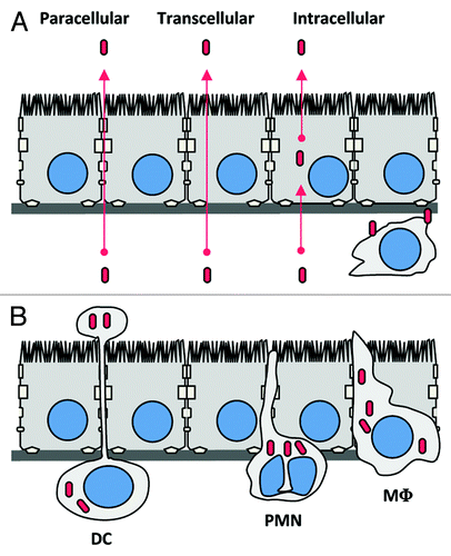
Figure 3. Potential mechanisms by which Lm could cross the blood-brain barrier. (A) Extracellular Lm, either free in the blood and/or associated to circulating cells, may recognize receptors at the surface of the barriers (as InlA, InlB or Vip) and cross them. (B) Trojan horse mechanism: Circulating leucocytes infected by Lm, such as monocytes, dendritic cells or polymorphonuclear cells, may cross the BBB hence targeting the bacteria in the CNS. DC, dendritic cell; PMN, polymorphonuclear leukocyte; M?, monocyte/macrophage.