1,832
Views
35
CrossRef citations to date
0
Altmetric
Review

Blood-brain barrier and retroviral infections

, , &
Pages 222-229 | Published online: 01 Mar 2012

Figures & data

Figure 1. Transmission electron microscopy (A) and schematic view (B) of rat brain illustrating the neurovascular unit. This complex includes microvessel endothelial cells (EC), based on basal lamina (BL), astrocytes end-feet (AEF) and some neurons (N) in the vicinity. Scale bar: 0.5 μm.
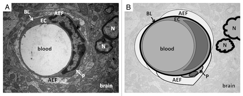
Figure 1. Transmission electron microscopy (A) and schematic view (B) of rat brain illustrating the neurovascular unit. This complex includes microvessel endothelial cells (EC), based on basal lamina (BL), astrocytes end-feet (AEF) and some neurons (N) in the vicinity. Scale bar: 0.5 μm.

Figure 2. Possible mechanisms of blood-brain barrier disruption by HTLV-1-infected lymphocytes. During TSP/HAM, HTLV-1 infected lymphocytes may disrupt the blood-brain barrier either by proinflammatory cytokine secretion (TNF-α, IL-1α) that activate NFκB pathway in endothelial cells, which induce tight junction disruption, or by infecting endothelial cells; the viral protein Tax could then induce tight junction disorganization.
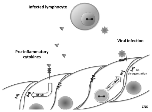
Figure 2. Possible mechanisms of blood-brain barrier disruption by HTLV-1-infected lymphocytes. During TSP/HAM, HTLV-1 infected lymphocytes may disrupt the blood-brain barrier either by proinflammatory cytokine secretion (TNF-α, IL-1α) that activate NFκB pathway in endothelial cells, which induce tight junction disruption, or by infecting endothelial cells; the viral protein Tax could then induce tight junction disorganization.