Figures & data
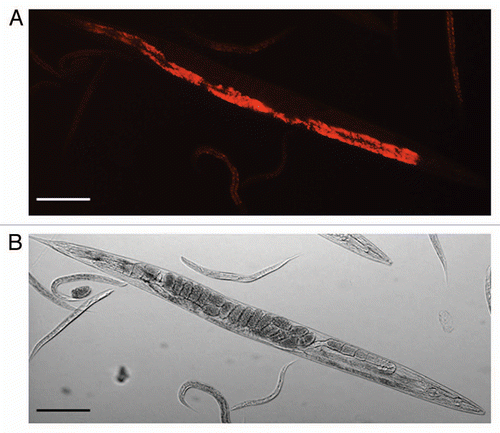
Figure 1 Light microscopic pictures of Caenorhabditis briggsae KT0001. (A) Red Fluorescent Protein (RFP)-tagged Serratia sp. SCBI lining the gut of C. briggsae KT0001. We used plasmid pSPR that harbors the red fluorescent protein gene from DsRedExpress for fluorescent tagging. For a detailed description of the methodology see reference Citation25. (B) Differential Interference Contrast image of same worm used in (A). (Scale bar = 100µm).
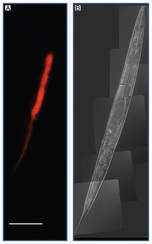
Figure 2 Light microscopic pictures of Caenorhabditis briggsae KT0001. (A) Red Fluorescent Protein (RFP)-tagged Serratia sp. SCBI lining the gut of C. briggsae KT0001. (B) Bright field image of same worm used in (A) (Scale bar = 100µm).