753
Views
20
CrossRef citations to date
0
Altmetric
Research Paper

Aspergillus terreus accessory conidia are multinucleated, hyperpolarizing structures that display differential dectin staining and can induce heightened inflammatory responses in a pulmonary model of aspergillosis

, , , , , , & show all
Pages 200-207 | Received 14 Jan 2011, Accepted 12 Apr 2011, Published online: 01 May 2011

Figures & data

Figure 1 Differential dectin-1 binding on Aspergillus fumigatus and Aspergillus terreus phialidic conidia. Soluble dectin-1 binding A. fumigatus and A. terreus phialidic conidia at various stages of germination (A) dormant conidia (B) swollen condia (C) early germ tube formation and (D) late germination. Differential interference microscopy (DIC) and fluorescence images were captured by microscopy at 40x (A) and 100x (B–D), and are representative of 3 experiments. Scale bars denote 5 µm and 10 µm for 40x and 100x magnification, respectively.
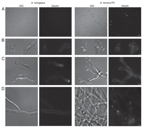
Figure 1 Differential dectin-1 binding on Aspergillus fumigatus and Aspergillus terreus phialidic conidia. Soluble dectin-1 binding A. fumigatus and A. terreus phialidic conidia at various stages of germination (A) dormant conidia (B) swollen condia (C) early germ tube formation and (D) late germination. Differential interference microscopy (DIC) and fluorescence images were captured by microscopy at 40x (A) and 100x (B–D), and are representative of 3 experiments. Scale bars denote 5 µm and 10 µm for 40x and 100x magnification, respectively.

Figure 2 Dectin-1 binding on phialidic conidia and accessory conidia at different germination stages. Soluble dectin-1 binding on A. terreus phialidic and accessory conidia at (A) dormant conidia (B) swollen conidia, (C) early germ tube formation and (D) late germination. DIC and fluorescence images were captured by microscopy at 40x (A) and 100x (B–D), and are representative of 3 experiments. Within windows, arrows depict the ring-like staining pattern on AC at 100x (A). Scale bars denote 5 µm and 10 µm for 40x and 100x magnification, respectively.
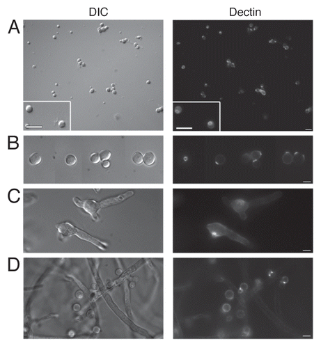
Figure 2 Dectin-1 binding on phialidic conidia and accessory conidia at different germination stages. Soluble dectin-1 binding on A. terreus phialidic and accessory conidia at (A) dormant conidia (B) swollen conidia, (C) early germ tube formation and (D) late germination. DIC and fluorescence images were captured by microscopy at 40x (A) and 100x (B–D), and are representative of 3 experiments. Within windows, arrows depict the ring-like staining pattern on AC at 100x (A). Scale bars denote 5 µm and 10 µm for 40x and 100x magnification, respectively.

Figure 3 Accessory conidia are multinucleated prior to germination. Hoechst staining was performed on A. terreus accessory conidia, both attached (A) and detached (B) from the hyphae, and A. terreus PC (C) and A. fumigatus PC (D). DIC and UV images were captured by microscopy at 100x, and are representative of 3 experiments. Scale bars denote 10 µm.
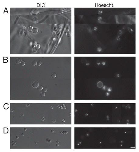
Figure 3 Accessory conidia are multinucleated prior to germination. Hoechst staining was performed on A. terreus accessory conidia, both attached (A) and detached (B) from the hyphae, and A. terreus PC (C) and A. fumigatus PC (D). DIC and UV images were captured by microscopy at 100x, and are representative of 3 experiments. Scale bars denote 10 µm.
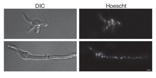
Figure 4 Accessory conidia undergo hyperpolarization during germination. Early germ tube formation and Hoechst nuclei staining was assessed for A. terreus accessory conidia. DIC and UV images were captured by microscopy at 100x, and are representative of 3 experiments. Scale bars denote 10 µm.

Figure 4 Accessory conidia undergo hyperpolarization during germination. Early germ tube formation and Hoechst nuclei staining was assessed for A. terreus accessory conidia. DIC and UV images were captured by microscopy at 100x, and are representative of 3 experiments. Scale bars denote 10 µm.

Figure 5 Aspergillus terreus accessory conidia elicit a heightened inflammatory response by alveolar macrophages. Alveolar macrophages were co-cultured with A. terreus AC or PC. Supernatants were collected at (A) 6 or (B) 20 h and assayed for chemokine/cytokine levels by Bio-Plex or ELISA. Experiments were performed three times.
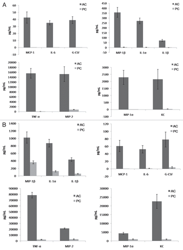
Figure 5 Aspergillus terreus accessory conidia elicit a heightened inflammatory response by alveolar macrophages. Alveolar macrophages were co-cultured with A. terreus AC or PC. Supernatants were collected at (A) 6 or (B) 20 h and assayed for chemokine/cytokine levels by Bio-Plex or ELISA. Experiments were performed three times.

Figure 6 Mice intratracheally challenged with Aspergillus terreus accessory conidia exhibit enhanced chemokine/cytokine production and cell recruitment in lungs. C57BL/6 mice were intratracheally challenged with AC or PC. (A) Chemokine/cytokine levels in the supernatants of homogenized lungs collected at 18 h post-challenge with AC or PC (need to indicate from figure). (B) Hematoxylin and eosin staining of sections of lungs post-challenge with AC or PC (also denote figure labels). All experiments were performed 3 times.
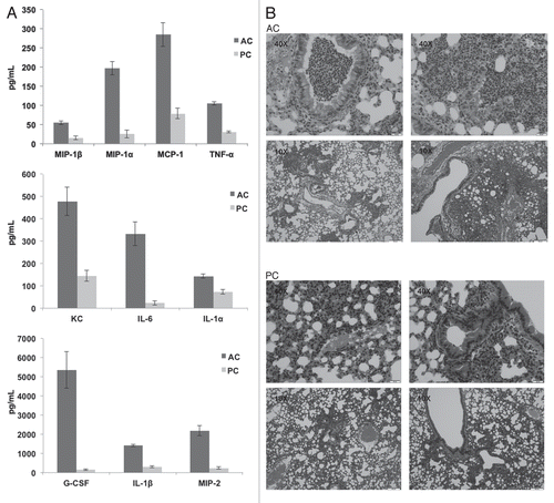
Figure 6 Mice intratracheally challenged with Aspergillus terreus accessory conidia exhibit enhanced chemokine/cytokine production and cell recruitment in lungs. C57BL/6 mice were intratracheally challenged with AC or PC. (A) Chemokine/cytokine levels in the supernatants of homogenized lungs collected at 18 h post-challenge with AC or PC (need to indicate from figure). (B) Hematoxylin and eosin staining of sections of lungs post-challenge with AC or PC (also denote figure labels). All experiments were performed 3 times.