Figures & data
Figure 1 Melanization dose response of C. neoformans to L-DOPA and Norepinephrine. Cells of strains JEC 21 (top row) and H99 (bottom row) were serially diluted and plated on media with or without the indicated concentration of substrate (mM). Plates were incubated for four (L-DOPA) or seven (Norepinephrine) days before photographing.
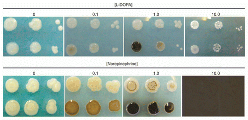
Figure 2 Effect of culture age on the rate of L-DOPA-mediated melanization. “New” indicates L-DOPA was added to cultures at the time of inoculation and incubated two days. “Old” indicates L-DOPA was added to a seven-day culture of C. neoformans and incubated for one or two more days. Left tube, strain JEC 21; right tube, strain H99.
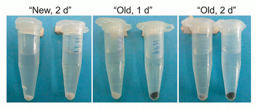
Figure 3 Effect of cell density on melanization. (A) C. neoformans strain JEC 21 was grown overnight in chemically defined minimal medium and concentrated before adding L-DOPA. Flasks were photographed after overnight incubation with L-DOPA. Cell densities of the cultures before adding L-DOPA were 6.6 × 10⁵, 4.6 × 10⁶ and 3.6 × 10⁷ CFU/ml. (B) L-DOPA agar was inoculated with 12, 135 or 1,250 CFUs of C. neoformans strain JEC 21 (top) or 14, 90 and 1,400 CFU of strain H99 (bottom) and incubated seven days at 30°C.
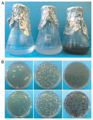
Figure 4 Fold changes in gene expression over time. C. neoformans wild-type (top) and laccase deletion (bottom) cultures were incubated with or without L-DOPA for 1–8 days. RNA was isolated from the cells at the indicated times for gene expression analysis by real-time PC R. The experiment was repeated twice. The results of one representative time course are shown. The fold changes in gene expression in the presence of L-DOPA are indicated for each gene.
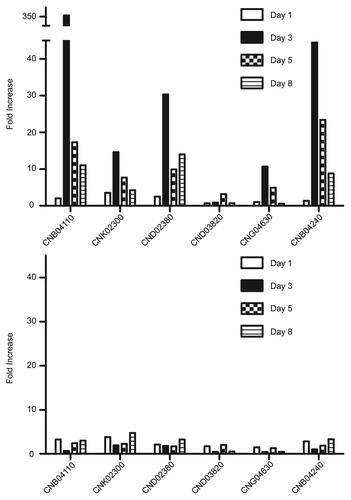
Figure 5 Incorporation of L-DOPA by cells. Assays were performed as described in the Methods and the percentage of L-DOPA incorporated calculated at indicated time intervals. Strains used were JEC 21 (WT), 2E-TU (Δlac1), 2E-TUC (COMPL.) and heated killed JEC 21 (HK).
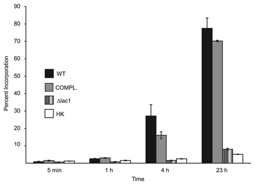
Table 1 Primers used for real-time PCR amplification
Table 2 Fold changes in gene expression upon addition of L-DOPA to cultures