Figures & data
Figure 1. LPS treatment causes a transient increase in VAMP3 mRNA expression. Expression of VAMP3 mRNA was analyzed in J774.A1 cells treated with either S. enterica serotype Minnesota LPS (SeM) or B. melitensis LPS. J774.1 cell were incubated for 30 or 60 min at 37°C in the presence of 200 ng/ml of SeM LPS or B. melitensis LPS. VAMP3 mRNA was quantified as described in Materials and Methods. Values are expressed in expression relative units (ERU). Bars represent the fold increase compared with its respective control cells at the same time points, by the Pfaffl equation. The results are representative of three independent experiments conducted in triplicate.
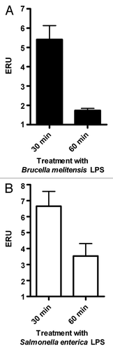
Figure 2. Infection by B. melitensis increases VAMP3 expression in J774.A1 macrophtages. J774.A1 cells were infected with B. melitensis (solid bars) or S. enterica serotype Enteritidis (SeE) (open bars) at a MOI of 50:1, at 37°C. After 15, 30, 45 and 60 min cultures were washed and cells harvested for RNA purification. VAMP3 mRNA was quantified as described in Materials and Methods. Values are expressed in expression relative units (ERU). Bars represent the fold increase compared with respective control cells at the same time points, by the Pfaffl equation. These results are representative of three independent experiments conducted in triplicate.
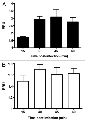
Figure 3. Expression of VAMP3 in J774.A1 cells treated with siRNA VAMP3. (A) qRT-PCR was performed at 6 h intervals to determine the time lapse in which VAMP3 is inhibited at maximum. Control corresponds to siRNA negative in western blotting. (B) Western blotting assay for VAMP3 of J774.A1 cells treated with siRNA VAMP3, samples were monitored each 2 h in order to detect maximum inhibition more precisely.
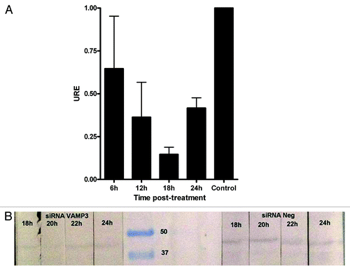
Figure 4. siRNA silencing of VAMP3 expression does not affect B. melitensis replication and survival in J774.A1 macrophages. J774.A1 cells transfected with three different siRNAs designed to silence mouse VAMP3 were infected with B. melitensis at a MOI of 50:1. The CFU counts, expressed as survival %, were determined at different intervals during a period of 36 h. Viable intracellular bacteria were determined by the gentamicin protection assay. Time zero represents total CFU counts previous to gentamicin treatment. The graph shows the mean of CFU counts from three independent experiments. *At this time point, a significant difference (p ≤ 0.05) was determined.
