1,603
Views
55
CrossRef citations to date
0
Altmetric
Research Paper

Galleria mellonella as a model host to study Paracoccidioides lutzii and Histoplasma capsulatum

, , , , &
Pages 139-146 | Received 17 May 2012, Accepted 28 Nov 2012, Published online: 09 Jan 2013

Figures & data

Figure 1. Log-Rank plots of the survival of G. mellonella after infection with different concentrations of P. lutzii yeast cells. G. mellonella infected and incubated at (A) 25°C or (B) 37°C. Controls included uninfected larva (Sham) and larva injected with PBS. n = 40 larvae per group.

Figure 1. Log-Rank plots of the survival of G. mellonella after infection with different concentrations of P. lutzii yeast cells. G. mellonella infected and incubated at (A) 25°C or (B) 37°C. Controls included uninfected larva (Sham) and larva injected with PBS. n = 40 larvae per group.

Figure 2. Log-Rank plots of the survival of G. mellonella after infection with different concentrations of H. capsulatum G184AR yeast cells. G. mellonella infected and incubated at (A) 25°C or (B) 37°C. Controls included uninfected larva (Sham) and larva injected with PBS. n = 60 larvae per group.

Figure 2. Log-Rank plots of the survival of G. mellonella after infection with different concentrations of H. capsulatum G184AR yeast cells. G. mellonella infected and incubated at (A) 25°C or (B) 37°C. Controls included uninfected larva (Sham) and larva injected with PBS. n = 60 larvae per group.

Figure 3. Log-Rank plots of the survival of G. mellonella after infection with different concentrations of H. capsulatum ATCC G217B yeast cells. G. mellonella infected and incubated at (A) 25°C or (B) 37°C. Controls included uninfected larva (Sham) and larva injected with PBS. n = 32 larvae per group.

Figure 3. Log-Rank plots of the survival of G. mellonella after infection with different concentrations of H. capsulatum ATCC G217B yeast cells. G. mellonella infected and incubated at (A) 25°C or (B) 37°C. Controls included uninfected larva (Sham) and larva injected with PBS. n = 32 larvae per group.

Figure 4. Infection of G. mellonella with H. capsulatum G184AR yeast cells induces melanization of the larva in a dose dependent manner. Larvae were injected with (A) PBS, (B) 101, (C) 102, (D) 103, (E) 104, (F) 105 or (G) 106 colonies of H. capsulatum/larvae. The images were taken 6 h after infection at 25°C.

Figure 4. Infection of G. mellonella with H. capsulatum G184AR yeast cells induces melanization of the larva in a dose dependent manner. Larvae were injected with (A) PBS, (B) 101, (C) 102, (D) 103, (E) 104, (F) 105 or (G) 106 colonies of H. capsulatum/larvae. The images were taken 6 h after infection at 25°C.

Figure 5. Infection of G. mellonella with H. capsulatum G184AR yeast cells induces melanization of the larva in a dose dependent manner. Larvae were injected with (A) PBS, (B) 101, (C) 102, (D) 103, (E) 104, (F) 105, or (G) 106 colonies of H. capsulatum/larvae. The images were taken 6 h after infection at 37°C.

Figure 5. Infection of G. mellonella with H. capsulatum G184AR yeast cells induces melanization of the larva in a dose dependent manner. Larvae were injected with (A) PBS, (B) 101, (C) 102, (D) 103, (E) 104, (F) 105, or (G) 106 colonies of H. capsulatum/larvae. The images were taken 6 h after infection at 37°C.

Figure 6. PAS-stained sections of G. mellonella. (A) Uninfected control larva inoculated with 0.1 M PBS. Larva infected with P. lutzii at concentrations of (B) 101, (C) 102, (D) 103, (E) 104 and (F) 105 colony forming units. All larva were incubated at 25°C. Structures are annotated as follows: a, adipose bodies; b, fungal cells; c, cuticle; h, hemolymph.
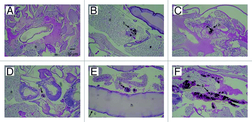
Figure 6. PAS-stained sections of G. mellonella. (A) Uninfected control larva inoculated with 0.1 M PBS. Larva infected with P. lutzii at concentrations of (B) 101, (C) 102, (D) 103, (E) 104 and (F) 105 colony forming units. All larva were incubated at 25°C. Structures are annotated as follows: a, adipose bodies; b, fungal cells; c, cuticle; h, hemolymph.
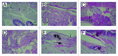
Figure 7. PAS-stained sections of G. mellonella. (A) Uninfected control larva inoculated with 0.1 M PBS. Larva infected with P. lutzii at concentrations of (B) 101, (C) 102, (D) 103, (E) 104 and (F) 105 colony forming units. All larva were incubated at 37°C. Structures are annotated as follows: a, adipose bodies; b, fungal cells; c, cuticle; h, hemolymph.

Figure 7. PAS-stained sections of G. mellonella. (A) Uninfected control larva inoculated with 0.1 M PBS. Larva infected with P. lutzii at concentrations of (B) 101, (C) 102, (D) 103, (E) 104 and (F) 105 colony forming units. All larva were incubated at 37°C. Structures are annotated as follows: a, adipose bodies; b, fungal cells; c, cuticle; h, hemolymph.

Figure 8. Number of granulomas containing yeast cells of P. lutzii as visualized microscopically on histology slides. PAS-stained sections of G. mellonella were counted using Image-Pro-Plus. The graphs represent the infected insects incubated at 25°C or 37°C. The control shown is insects injected with PBS. **p = 0.0027 comparing two temperatures.
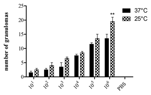
Figure 8. Number of granulomas containing yeast cells of P. lutzii as visualized microscopically on histology slides. PAS-stained sections of G. mellonella were counted using Image-Pro-Plus. The graphs represent the infected insects incubated at 25°C or 37°C. The control shown is insects injected with PBS. **p = 0.0027 comparing two temperatures.

Reprints and Corporate Permissions

Please note: Selecting permissions does not provide access to the full text of the article, please see our help page How do I view content?

To request a reprint or corporate permissions for this article, please click on the relevant link below:

Academic Permissions

Please note: Selecting permissions does not provide access to the full text of the article, please see our help page How do I view content?

Obtain permissions instantly via Rightslink by clicking on the button below:

If you are unable to obtain permissions via Rightslink, please complete and submit this Permissions form. For more information, please visit our Permissions help page.