906
Views
11
CrossRef citations to date
0
Altmetric
Brief Report

Constant domains influence binding of mouse–human chimeric antibodies to the capsular polypeptide of Bacillus anthracis

, , &
Pages 483-488 | Received 23 May 2013, Accepted 10 Jul 2013, Published online: 17 Jul 2013

Figures & data

Figure 1. Binding of anti-capsular antibodies to PGA-coated sensor surface. The total RU that was achieved following a 180 sec pulse is given as a function of antibody concentration. The KD of each antibody was determined using the steady-state equilibrium model in BIAevaluation software, where the theoretical RUmax is determined and the apparent KD is equal to the antibody concentration that is necessary to reach ½ RUmax.
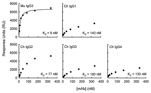
Figure 1. Binding of anti-capsular antibodies to PGA-coated sensor surface. The total RU that was achieved following a 180 sec pulse is given as a function of antibody concentration. The KD of each antibody was determined using the steady-state equilibrium model in BIAevaluation software, where the theoretical RUmax is determined and the apparent KD is equal to the antibody concentration that is necessary to reach ½ RUmax.

Table 1. Immunochemical activities of murine and mouse–human anti-PGA F26G3 antibodies

Figure 2. Capsule reaction produced by antibody binding. Top panel: Visualization of capsular polypeptide following the addition of F26G3 MuAbs IgG3 or PBS. Addition of the F26G3 MuAb IgG3 (100–150 μg/mL) produces a rim-type reaction with deposition both proximal to the cell wall and at the capsular edge. The capsule is not visible in the absence of specific antibody (PBS). Bottom panel: Visualization of capsular polypeptide following the addition of F26G3 ChAbs IgG1–4 (100 μg/mL). The addition of F26G3 ChAbs produces a puffy-type reaction that occurs uniformly throughout the capsule.
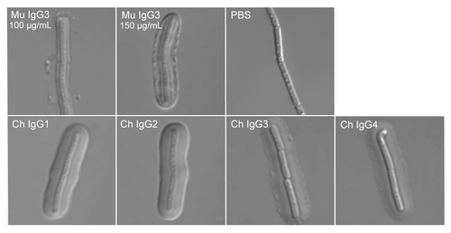
Figure 2. Capsule reaction produced by antibody binding. Top panel: Visualization of capsular polypeptide following the addition of F26G3 MuAbs IgG3 or PBS. Addition of the F26G3 MuAb IgG3 (100–150 μg/mL) produces a rim-type reaction with deposition both proximal to the cell wall and at the capsular edge. The capsule is not visible in the absence of specific antibody (PBS). Bottom panel: Visualization of capsular polypeptide following the addition of F26G3 ChAbs IgG1–4 (100 μg/mL). The addition of F26G3 ChAbs produces a puffy-type reaction that occurs uniformly throughout the capsule.

Reprints and Corporate Permissions

Please note: Selecting permissions does not provide access to the full text of the article, please see our help page How do I view content?

To request a reprint or corporate permissions for this article, please click on the relevant link below:

Academic Permissions

Please note: Selecting permissions does not provide access to the full text of the article, please see our help page How do I view content?

Obtain permissions instantly via Rightslink by clicking on the button below:

If you are unable to obtain permissions via Rightslink, please complete and submit this Permissions form. For more information, please visit our Permissions help page.