Figures & data
Figure 1. Characterization of purified TLF-1 and TLF-2. (A) Superose 6 size exclusion chromatography of TLF-1 and TLF-2. Absorbance profiles (280 nM) of TLF-1 and TLF-2, superimposed on individually ran marker proteins [1, thyroglobulin (660 kDa); 2, apoferritin (480 kDa); 3, conalbumin (67 kDa); 4, ovalbumin (45 kDa)]. (B) Analysis of individual Superose 6 column fractions of TLF-1 and TLF-2 separated on non-denaturing 12% SDS-PAGE and silver stained (top panel). Hpr, apoL1 and IgM were detected by protein gel blot. NA, not analyzed.
![Figure 1. Characterization of purified TLF-1 and TLF-2. (A) Superose 6 size exclusion chromatography of TLF-1 and TLF-2. Absorbance profiles (280 nM) of TLF-1 and TLF-2, superimposed on individually ran marker proteins [1, thyroglobulin (660 kDa); 2, apoferritin (480 kDa); 3, conalbumin (67 kDa); 4, ovalbumin (45 kDa)]. (B) Analysis of individual Superose 6 column fractions of TLF-1 and TLF-2 separated on non-denaturing 12% SDS-PAGE and silver stained (top panel). Hpr, apoL1 and IgM were detected by protein gel blot. NA, not analyzed.](/cms/asset/17ab197f-2f65-45a6-aad2-227782a58cd0/kvir_a_10918295_f0001.gif)
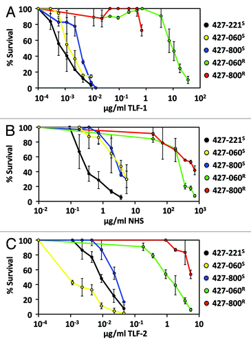
Figure 2. In vitro activity of human serum, TLF-1 and TLF-2. TLF-1 resistant (R) and susceptible (S) clonal cell lines of bloodstream form T. b. brucei Lister 427 expressing VSG221, 800 and 060 were prepared as previously described.Citation24,Citation25 The percentage surviving cells was determined, using phase contrast microscopy, 14 h following the addition of TLF-1, TLF-2 or complete human serum to exponentially growing cultures at 37°C. (A) TLF-1 susceptibility of T. b. brucei 427-221S (black), T. b. brucei 427-800S (blue), T. b. brucei 427-800R (red), T. b. brucei 427-060S (yellow) and 427-060R (green). (B) Normal human serum (NHS) susceptibility of T. b. brucei 427-221S (black), T. b. brucei 427-800S (blue), T. b. brucei 427-800R (red), T. b. brucei 427-060S (yellow) and 427-060R (green). (C) TLF-2 susceptibility of T. b. brucei 427-221S (black), T. b. brucei 427-800S (blue), T. b. brucei 427-800R (red), T. b. brucei 427-060S (yellow) and 427-060R (green).