

Abstract
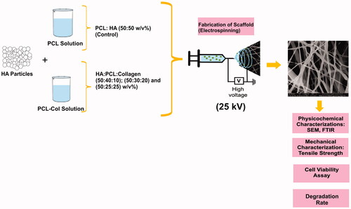
According to the Indonesian Ministry of Health Research and Development Agency's 2018 Basic Health Research (Riskesdas), bone defects, because of fracture cases in Indonesia, reached a prevalence of 5.5%. Bone tissue engineering is one of the most promising new approaches for accelerating the growth and healing of bone defects in patients. In this study, a bone scaffold electrospun nanofiber composed of hydroxyapatite (HA)-polycaprolactone (PCL)-collagen was fabricated to mimic the extracellular matrix (ECM) found in native bone tissue and to promote bone remodeling and healing. Through characterization of functional groups with Fourier transform infrared (FTIR), morphology scanning electron microscopy (SEM), tensile strength test, degradation test, and cytotoxicity MTT (3-(4,5-Dimethylthiazol-2-yl)-2,5-diphenyltetrazolium bromide) assay, this study determines the optimal composition of electrospun nanofiber HA/PCL/collagen to obtain the best candidate of a bone scaffold with excellent characteristics. Electrospinning was used to fabricate bone scaffolds. The samples used in this study consisted of control samples, namely PCL-HA and PCL-Collagen, and three samples with a mass ratio composition of HA-PCL-collagen 50:40:10, 50:30:20, and 50:25:25 (w/v%). The FTIR analysis of the sample revealed that no chemical bond existed between the materials. The HA/PCL/collagen 50:25:25 (F3) sample exhibited the best characteristics as a bone scaffold candidate, with a fiber diameter of 365 ± 202 nm; a porous fraction area of 58.98%; an ultimate tensile strength, and an elasticity modulus of 0.60 ± 0.185 and 5.98 ± 0.82 MPa, respectively; a degradation rate of 1.93 × 10⁻⁶ g/h; and cell viability of 81.03%.
Graphical Abstract
Acknowledgments
The authors also would like to acknowledge the use of facilities and technical assistance from the Biomedical Technology and Material Physics Laboratories, Faculty of Science and Technology, Universitas Airlangga, and the Material Physics and Electronics Laboratory, as well as the staff of the Integrated Laboratory for Research and Testing at the Universitas Gadjah Mada, Indonesia.
Disclosure statement
No potential conflict of interest was reported by the author(s).
Data availability statement
The datasets used and/or analyzed during the current study are available from the corresponding author on reasonable request.