
Abstract
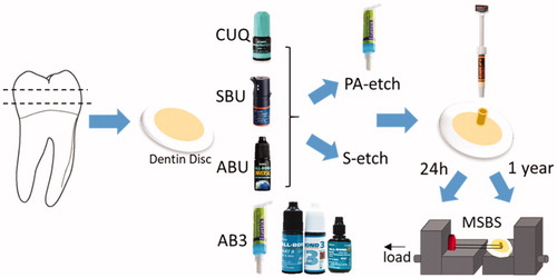
This study aimed to compare the microshear bond strength (MSBS) of three universal adhesives and a three-step conventional adhesive to dentin after 24-hour and one-year storage in water. A new fluoride-releasing universal adhesive (Clearfil Universal Bond Quick: CUQ) and two commercially available adhesives (ScotchBond Universal: SBU and All-Bond Universal: ABU) were evaluated with phosphoric acid etching (PA-etch mode) or without it (self-etch mode). All-Bond 3 (AB3) served as control group. After bonding composite cylinders to dentin discs obtained from caries-free human teeth, the specimens were stored in deionized water at 37 °C for either 24 hours or one year (n = 14) before MSBS measurement. Two-way ANOVA analysis of the results showed that the adhesives, storage time and their interactions had a significant effect on MSBS (p < 0.01). In self-etch mode, there was no significant difference among universal adhesives at the baseline. In PA-etch, the CUQ and SBU showed significantly higher MSBS compared with AB3 (p < 0.05). At baseline, no difference was found between the two modes for each universal adhesive (p > 0.05). After one year, CUQ in self-etch mode showed a slight increase in nominal MSBS (p > 0.05) and Weibull characteristic strength, which was significantly higher than SBU and ABU in the corresponding mode. There was no difference among the three universal adhesives in PA-etch mode after one year (p > 0.05). In conclusion, the durability and reliability of dentin bonding with universal adhesives in different application modes depended on the material; and the self-etch approach showed promising results for the tested fluoride-releasing universal adhesive.
1. Introduction
Tooth-colored restorations have become increasingly popular with the development of adhesive dentistry. Dental adhesives provide bonding of composite resin to the dental hard tissue, mainly enamel and dentin. Good and durable bonding to enamel was established several decades ago [Citation1], however, bonding to dentin has been historically facing more challenges [Citation2].
Dentin adhesive systems traditionally relied on three application steps: phosphoric acid (PA) etching to remove the smear layer, open dentin tubule orifices and demineralize dentin matrix followed by rinsing; application of a hydrophilic primer to re-expand the organic matrix and facilitate penetration of the bonding resin; and finally bonding, which comprises application of a resin to penetrate the primed dentin and polymerize to form adhesion. Self-etch systems were later developed based on the functional monomer chemistry [Citation3], which could simultaneously demineralize dentin superficially and penetrate the resin monomers. The self-etch adhesives can reduce clinical operation time and minimize the technique sensitivity [Citation4]. Earlier self-etch adhesive systems required a separate bonding agent application after the self-etch primer; more recently all-in-one adhesives have been introduced as simplified adhesives in one-bottle. However, the effectiveness of the simplified steps of bonding has been controversial. It has been reported that the bonding performance of all-in-one adhesives are inferior to that of both the 3-step and 2-step self-etching systems and have a higher annual failure compared to etch-and-rinse adhesives [Citation5–7].
The universal adhesives appeared as the latest generation of bonding agents that can perform with or without PA-etch for enamel and dentin [Citation8–11]. Universal adhesives are designed under the all-in-one concept of the existing one-step self-etch adhesives and incorporate the versatility for clinical situations. According to the manufacturers of universal adhesives, the clinician can decide between etch-and-rinse or self-etch mode for enamel and dentin. While there is increasing evidence that PA-etch benefits bonding of universal adhesives to enamel, the same consensus has not been reached for dentin. Some earlier works suggested that etching may have detrimental effects on the longevity of resin-dentin bond in the long-term [Citation8].
In single-step all-in-one adhesives including universal adhesives, the hydrophilic and hydrophobic monomers are blended with water and solvents that may create problems with regard to the homogeneity of the blend or long term degradation of bond [Citation4,Citation9,Citation12–14]. Simplified procedures and elimination of cross-contamination risks are great benefits of the all-in-one adhesive, however, long-term durability and necessity of PA-etch step on dentin need further clarification [Citation9,Citation15].
While the initial strength is essential for evaluation of an adhesive, durability also plays an important role for the longevity of a restoration. Some researchers reported that a critical decrease in bond strength after long-term usage may reduce the success rate of restorations and lead to loss of retention, leakage and other failure [Citation12,Citation16]. It is still important to investigate the durability of adhesive bonds to dentin with the newly introduced universal adhesives [Citation11,Citation12,Citation17,Citation18].
The aim of this study was to evaluate the microshear bond strength (MSBS) of universal adhesives to dentin with or without PA-etch after one-year storage in water. The null hypotheses of the study were (1) there was no difference in MSBS after one-year storage in water compared to the baseline; and (2) MSBS of universal adhesives was not affected by the PA-etch.
2. Materials and methods
2.1. Restorative materials used
Four current adhesives were evaluated in this study. Firstly, three universal adhesives including experimental universal adhesive Clearfil Universal Bond Quick (CUQ), ScotchBond Universal adhesive (SBU) and Bisco All-Bond universal (ABU) were used. They were applied in two modes, with and without PA etching, namely total or etch-and-rinse method and all-in-one step self-etch method, respectively. Secondly, a three-step total-etch adhesive, Bisco All-Bond 3 (AB3) were utilized. The composite Clearfil AP-X was used to evaluate the MSBS of the adhesives to dentin. The adhesives used in this study and their compositions were listed in .
Table 1. List of materials used.
2.2. Sample preparation
The methodology of the study was illustrated in . The usage of human teeth were subjected to the guideline of the Ethic Committee of University of Washington in accordance with the principles of the Declaration of Helsinki. More than eighty freshly extracted human noncarious premolars and molars were collected, stored at 4 °C in water with 0.02% thymol. Forty teeth were used for MSBS tests.
Figure 1. Schematic representation of study design. Microshear bond strength of adhesives to human dentin was measured 24 hour and one year after bonding with different adhesives.
The occlusal cusps were removed to expose the dentin using a cutting machine (Sankei aqra preciso CL-40, Japan) with a low-speed diamond saw at the speed of 40 rpm under water cooling. The roots were subsequently cut at the cement-enamel junction. 96 dentin slices of 2-mm-thickness were obtained. The surface of the superficial dentin slices were polished under running water by 600 grit silicone paper (3 M ESPE, USA) to create a standardized smear layer. The surfaces were checked under a microscope digital camera (AmScope MU1000, USA) to exclude the cracks, residual enamels, coarseness and contamination.
The dentin disks were randomly distributed into 7 groups according to the adhesives and application modes. The application of the materials was performed according to the manufacturer’s instructions. For dentin etching with PA, specimens were conditioned with the acid gel for 15 s, thoroughly rinsed with water, blotted using a cotton pellet to remove remaining water. They were left visibly moist before the bonding agent was applied. For adhesive application, the dentin surface was coated with a layer of bond of using the microbrushes supplied by the manufacturer. The surfaces were gently air-dried to spread the adhesive evenly and evaporate the solvents.
2.3. Microshear bond test
Prior to the irradiation (light curing), two or three tygon tubes (Saint-Gobain Performance Pastic, Nagano, Japan) with an internal diameter of 0.79 mm and 1.0 mm in height were placed on the dentin surface of each slice. The adhesive was light cured for 10 s at 600 mw/cm2 intensity using a halogen light-curing unit (Yoshida, Japan). The composite Clearfil AP-X was then carefully inserted into the tubing lumens and irradiated for 40 s. The specimens were stored at room temperature (23 °C) for 1 h then the tygon tubes were carefully removed using a sharp blade. In this manner, the small cylinders of resin approximately 0.79 mm in diameter and 1.0 mm in height remained bonded to the dentin surface. All the specimens were stored in distilled water in an incubator at 37 °C [Citation12,Citation13,Citation17].
Each experimental group was divided into two subgroups according to the storage time: 24 hours and one year (n = 14). MSBS was measured after storage for each group. The dentin slice was attached to the testing apparatus with a cyanoacrylate adhesive (Zapit, Dental Ventures of America Inc., USA) and tested in a compact tester (Bisco, Inc., Schaumburg, IL, USA). A thin steel wire (0.2 mm diameter) was looped at the bottom of the resin cylinder, in contact with the lower half-circle of the cylinder and the dentin surface. The wire loop was kept in the manner to maintain the shear stress orientation at the bonding interface. The force was applied by pulling the wire at a crosshead speed of approximately 1 mm/min until failure occurred and the MSBS data were immediately recorded [Citation18].
2.4. Evaluation of failure mode
After MSBS test, the mode of failure was determined for each specimen with a digital stereomicroscope (AmScope MU1000, Irvine, CA, USA). The fractured surfaces were classified according to the following criteria: adhesive failures between resin and dentin, mixed failures that were partially adhesive and partially cohesive, and cohesive failures that occurred entirely within the bonding resin or entirely in dentin [Citation19].
2.5. Scanning electron microscope (SEM)
The sample preparation for visualization under SEM (JEOL JSM-6010LA, Tokyo, Japan) was similar to that for the MSBS test. After air drying, the adhesive-coated surface was irradiated for 10 s. Composite Clearfil AP-X was placed as a build-up of 2 mm in thickness on the bonding surface and irradiated 40 s. After storing in distilled water at 37 °C for 24 h, the bonded specimens were cut perpendicular in the buccolingual direction by the diamond saw to obtain two halves. Each group obtained 2 dentin slices. The slices were embedded in epoxy resin (TotalBoat, Bristol, RI, USA). The section surfaces were polished to a high gloss using waterproof SiC paper from 600 grit to 2000 grit. Abrasive discs were used followed by diamond pastes from 6 µm down to 0.25 µm to create a smooth surface. Each specimen was dehydrated, subjected to argon-ion beam etching, gold sputter coated, and observed under the SEM [Citation17].
2.6. Statistical analysis
The MSBS values were statistically analyzed using two-way analysis of variance (ANOVA) was applied at a significance level of 0.05 with MSBS as dependent variable and the adhesive material and storage time as factors. In addition, the Weibull distribution parameters were determined for each group using the linear regression method at the 95% confidence level.
3. Results
3.1. Microshear bond strength
The results of MSBS test for each group (mean and SD) were presented in . MSBS values ranged 20 to 65 MPa. Two-way ANOVA with adhesive material and storage time as factors indicated that both factors and their interactions were significant (p < 0.01). In self-etch mode, there was no significant difference among universal adhesives at the baseline. In PA-etch, the CUQ and SBU showed significantly higher MSBS compared with AB3 (p < 0.05). ANOVA showed no significant difference between the two modes at baseline or after one-year within each universal adhesive (p > 0.05).
Table 2. Microshear bond strength Mean ± SD in MPa (n = 14).
MSBS values of CUQ in self-etch mode showed no significant difference between baseline and one-year storage (p > 0.05). The values in this group were higher than those of self-etch SBU and ABU after one year (p < 0.05). The values of SBU in PA-etch mode also showed no significant difference between baseline and one-year storage (p > 0.05). However, there was no difference between SBU, CUQ and ABU in PA-etch mode after one year (p > 0.05). Except for the self-etch CUQ and PA-etch SBU, all other groups showed significant reduction in MSBS after one year of storage (p < 0.05).
summarizes Weibull modulus (m) and 95% confidence interval (CI) of m, characteristic strength (σ0) and 95% confidence interval of σ, 0.5% probability of failure (σ0.05) and Weibull coefficient of correlation (r) for each of the experimental group. The data and Weibull distribution fitted with good correlation coefficient value. Weibull analysis showed that the characteristic strength of most groups decreased after one-year storage, when the probability curve shifted to the left, except for CUQ in self-etch mode () (). An evident drop was observed for σ0.05 when comparing bond strength after 24-hour and one-year storage for most groups except for CUQ. On the other hand, the Weibull modulus showed high scattering of data for CUQ in PA-etch mode, which improved and stabilized over the one-year storage.
Figure 2. Probability of failure vs. stress for the experimental groups at the baseline (BL) and after one-year (1Y) storage. The lines were drawn at 63% and 5% probability of failure: a) AB3; b) ABU; c) SBU; d) CUQ.
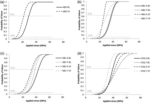
Table 3. Microshear bond strength Weibull statistics.
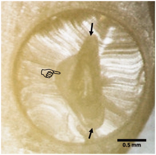
The failure mode observation suggested that the failures were predominantly mixed involving bonding resin and dentin interface, which is a typical observation for the wire-loop MSBS test ().
Figure 3. Typical MSBS mixed failure mode under stereomicroscope. The composite resin cylinder separation from the dentin has resulted in exposure of dentin surface (finger pointer) and partial fracture of the adhesive layer (arrows).
3.2. SEM images
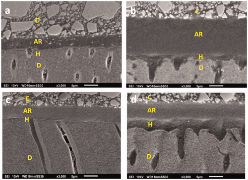
SEM images () suggested that the adhesive layer ranged 5 to 20 µm in thickness among universal adhesive groups. The low density interfacial zone (so-called hybrid layer) showed 3–5 µm thickness with PA-etch, but appeared as a thin zone (<1 µm thick) under the self-etch mode. Demineralization effects of PA created a serrated pattern, and wide-open dentinal tubules with tubular resin tag formation were apparent in PA-treated groups. However, in the self-etch mode, the dentin surface appeared smooth, smear plugs remained, perpendicular-cut tubules were sealed, and tubular resin tags were observed just occasionally.
Figure 4. Representative SEM images indicating the difference in adhesive layers of experimental groups at baseline; a) ABU-S; b) CUQ-PA; c) SBU-S; d) SBU-PA. The adhesive layer ranged 5 to 20 µm in thickness among universal adhesive groups. The low-density interfacial zone (so-called hybrid layer) showed 3–5 µm thickness with PA etching (b, d), but appeared as a thin zone (<1 µm thick) under the self-etch mode (a, c). Demineralization effects of PA, creating a serrated pattern and wide-open dentinal tubules with tubular resin tag formation were apparent in PA-treated groups (b, d). C: Composite; AR: Adhesive Resin; H: Hybrid Layer; D: Dentin.
4. Discussion
Based on the results of this study, a general recommendation may not be made with regard to the PA-etch prior to application of the universal adhesives. Consistent with other literatures, the dentin bond strength of universal adhesives was not evidently improved with PA-etch [Citation8,Citation11]. Regardless of the application mode, universal adhesives showed that they have reliable immediate bond strength to dentin, which is in line with previous reports [Citation20]. The MSBS in the groups, except the CUQ self-etch, somewhat decreased after one-year storage. The decrease was not statistically significant for SBU PA-etch. However, based on the ANOVA and Weibull, it is difficult to argue that there was a strong advantage with PA-etch for SBU.
The first hypothesis was rejected as the MSBS were different among the groups and the storage time affected the MSBS. The second hypothesis, which stated that MSBS of universal adhesives was not affected by the PA-etch was accepted, as there was no difference between the two modes within each adhesive at each time. All the universal adhesives showed better or comparable MSBS to that of the conventional three-step adhesive AB3.
In this study, the MSBS test was employed to assess the bond strength of universal adhesives in two modes of applications, with or without PA-etch. MSBS test is one of the popular methods for evaluating effectiveness of adhesives. It has been suggested that the MSBS test has the advantage of less time-consuming and better controlled specimen preparation [Citation21]. In order to investigate the bond strength reliability, Weibull analysis provides information of the reliability rather than solely relying on the average bond strength and standard deviation. This method allows the prediction of the likelihood of failure of a material at low stress values. It enables the clinician to select a material based on an expected level of stress that might apply on a restoration in oral cavity. Weibull modulus (m) values were in the range of adhesive materials from previous studies [Citation18]. The values are generally higher in baseline than one-year storage, denoting a higher scatter in bond strength results in long-term. The results are in alignment with previous work showing a decrease in the characteristic strength after aging using thermal cycling [Citation18]. The Weibull failure probability curves shifted to the left for all the tested adhesives in both modes with one-year storage, denoting an increased probability of failure at lower stress, with the exception of CUQ in self-etch mode which slightly shifted to the right. This concurred with the results of SBU, ABU in both modes and AB3 experienced a drop in σ 0.05, at the stress at 5% failure probability.
The compositions of the universal adhesives and the all-in-one are similar, since they both contain all hydrophilic components, including functional monomers providing the functions of etching and polymerization. The 10-Methacryloyloxydecyl dihydrogen phosphate (MDP) has received great attention over the past years as an adhesive dental monomer suitable for self-etch and universal formulations. MDP contains both hydrophilic and hydrophobic parts. Addition of MDP into a bonding resin may render characteristics of adhesive resin more hydrophilic. This acidic functional monomer contributes to increase the initial bond strength to dentin due to increased interaction with the substrate. The monomer has the potential to develop chemical bonds to dentin minerals; it has been reported that phosphate group of MDP has a potential to interact with hydroxyapatite and contributes significantly to the long-term durability of dentin-resin bonding interface [Citation3]. The lowest MSBS at baseline was shown in AB3 group, where MDP is absent in the formula of the conventional three-step adhesive. It appears that the functional monomer plays a key role in the improvement of bonding regardless of the application steps. On the other hand, some concerns have been mentioned with regard to MDP in the formulations; it would increase the water sorption capability of the cured adhesive in the long-term storage and decrease the overall degree of conversion in the polymer [Citation3].
Morphological SEM micrographs in this study suggested that more aggressive etching by PA resulted in thicker apparent hybrid layer and rougher dentin surface, which was expected. Hybrid layer and resin tag formation are dependent on the adhesive system, dentin surface characteristics, intertubular dentin, and tubule orientation [Citation6]. It is conceived that PA-etch removes the smear layer, promotes superficial dentin demineralization and degradation with the dentin [Citation10,Citation11,Citation22]. The use of PA dissolves some of the intratubular mineral deposits, creating more resin tags than compared to the self-etch mode. However, it was argued that the principal method of adhesion to the dentin may not depend on resin tag formation, but on chemical bonding to the hydroxyapatite [Citation10]. If this is the case, then PA pre-etching on dentin may extensively remove hydroxyapatite from the surface, which seems necessary to achieve chemical bonding with MDP [Citation23].
Under a milder demineralization, the preserved mineral (apatite) together with MDP served as the substrate of chemical interaction. This was consistent among the groups given the mild acidity of the universal formulations (pH > 2.0). As a result of self-etch mode, the acid-resistance of interface has been reported to improve and the organic phase of dentin remained intact [Citation23].
Some authors suggested that 20 µm was the minimum adhesive thickness to avoid polymerization inhibition by the oxygen [Citation24]. The adhesive layer in universal bonds in this study appeared to be 5 to 20 µm thick. The thinner the layer is, the higher chance that polymerization is inhibited. The ideal minimum thickness for the adhesive layer is still a controversial issue and deserves future investigations [Citation24, Citation25].
SBU-PA showed a gradient of density decreasing from the adhesive towards the dentin, while in CUQ-PA, a more homogenous zone appeared as a layer (). Hydroxyethyl methacrylate (HEMA) is a hydrophilic methacrylate that possesses the capability of increasing bond strength to dentin [Citation5,Citation26]. However, owing to its characteristics, it could enhance water uptake leading to swelling. The mechanical properties of the polymerized adhesive deteriorate, decreasing bond strength after long time water storage [Citation2,Citation22,Citation27,Citation28]. According to the manufacturer, CUQ has a new amide monomer [Citation25] that was intended to improve the penetration of adhesive to the tooth substrate and improve polymerization of the adhesive blend, which in turn results in less fluid absorption. The manufacturer also claims that as a result of improved penetration of the adhesive into dentin, the need for rubbing or agitation is eliminated with CUQ; however, in this study we followed the standard 20 s agitation time for all adhesives. It was reported that the performance of universal adhesives could be compromised in the application using a shortened application time [Citation29]. Prior to polymerization, the amide is much more hydrophilic than HEMA and can penetrate especially well into dentin. An important advantage of amide monomers is that acrylamide provides better hydrolytic resistance to either groups.
The degradation of bond over storage has been attributed to auto-degradation of collagen matrices which occur in resin-infiltrated dentin by the slow action of host-derived matrix metalloproteinases (MMPs) [Citation30,Citation31]. This degradation has been shown for both etch-and-rinse with PA and self-etch approaches; however, generally a higher level of enzymatic activity was seen following PA-etching compared with self-etching, which correlated with more rapid destruction of hybrid layers [Citation32]. This degradation was observed in characteristic strength of all groups, except for CUQ in self-etch mode.
The results of Weibull statistics are interesting with regard to the CUQ. In self-etch mode, the adhesive experiences an increase in the characteristic strength. A previous study reported similar findings with regard to the mean MSBS of a fluoride-releasing two-step self-etch adhesive [Citation33], and attributed the increase to the effects of fluoride release in terms of stabilizing the bonding of the MDP-based adhesive. These results are corroborated by other reports showing evidence of stable interaction of MDP with apatite in the presence of low level of fluoride [Citation34] and with all-in-one adhesive compared to non-fluoride releasing adhesive [Citation35]. Future studies will need to elaborate further on ion release from dental adhesives.
5. Conclusions
Within the limitations of this laboratory study, the bond strength of the universal adhesives was not affected by the mode of application. The tested universal adhesives showed higher bond strength than the conventional three-step adhesive to acid-etched dentin. The universal adhesive formulation containing hydrophilic amide monomer and sodium fluoride showed improved stability particularly in the self-etch mode.
Acknowledgements
The authors would like to thank Bisco Inc. and Kuraray Noritake Dental for their support to this research by donating the adhesive materials used. Authors are also thankful to Natasha Paranjapye and Selene Sarraf for their help.
Disclosure statement
No potential conflict of interest was reported by the authors.
References
- Buonocore MG. A simple method of increasing the adhesion of acrylic filling materials to enamel surfaces. J Dent Res. 1955;34:849–853.
- De Munck J, Mine A, Poitevin A, et al. Meta-analytical review of parameters involved in dentin bonding. J Dent Res. 2012;91:351–357.
- Matsui N, Takagaki T, Sadr A, et al. The role of MDP in a bonding resin of a two-step self-etching adhesive system. Dent Mater J. 2015;34:227–233.
- Giannini M, Makishi P, Ayres AP, et al. Self-etch adhesive systems: a literature review. Braz Dent J. 2015;26:3–10.
- Fu J, Pan F, Kakuda S, et al. The effect of air-blowing duration on all-in-one systems. Dent Mater J. 2012;31:1075–1081.
- Freeman R, Varanasi S, Meyers IA, et al. Effect of air abrasion and thermocycling on resin adaptation and shear bond strength to dentin for an etch-and-rinse and self-etch resin adhesive. Dent Mater J. 2012;31:180–188.
- Özcan E, Çetin AR, Tunçdemir AR, et al. The effect of luting cement thicknesses on the push-out bond strength of the fiber posts. Acta Odontol Scand. 2013;71:703–709.
- Rosa WL, Piva E, Silva AF. Bond strength of universal adhesives: a systematic review and meta-analysis. J Dent. 2015;43:765–776.
- Hanabusa M, Mine A, Kuboki T, et al. Bonding effectiveness of a new ‘multi-mode' adhesive to enamel and dentine. J Dent. 2012;40:475–484.
- Muñoz MA, Sezinando A, Luque-Martinez I, et al. Influence of a hydrophobic resin coating on the bonding efficacy of three universal adhesives. J Dent. 2014;42:595–602.
- Wagner A, Wendler M, Petschelt A, et al. Bonding performance of universal adhesives in different etching modes. J Dent. 2014;42:800–807.
- Sadr A, Shimada Y, Tagami J. Effects of solvent drying time on micro-shear bond strength and mechanical properties of two self-etching adhesive systems. Dent Mater. 2007;23:1114–1119.
- Sadr A, Ghasemi A, Shimada Y, et al. Effects of storage time and temperature on the properties of two self-etching systems. J Dent. 2007;35:218–225.
- Grégoire G, Dabsie F, Dieng-Sarr F, et al. Solvent composition of one-step self-etch adhesives and dentine wettability. J Dent. 2011;39:30–39.
- Vermelho PM, Reis AF, Ambrosano GM, et al. Adhesion of multimode adhesives to enamel and dentin after one year of water storage. Clin Oral Invest. 2016;21:1707–1715.
- El-Deeb HA, Badran O, Mobarak EH. One-year adhesive bond durability to coronal and radicular dentin under intrapulpal pressure simulation. Oper Dent. 2015;40:540–547.
- Wei S, Sadr A, Shimada Y, et al. Effect of caries-affected dentin hardness on the shear bond strength of current adhesives. J Adhes Dent. 2008;10:431–440.
- Hariri I, Shimada Y, Sadr A, et al. The effects of aging on shear bond strength and nanoleakage expression of an etch-and-rinse adhesive on human enamel and dentin. J Adhes Dent. 2012;14:235–243.
- Moosavi H, Hariri I, Sadr A, et al. Effects of curing mode and moisture on nanoindentation mechanical properties and bonding of a self-adhesive resin cement to pulp chamber floor. Dent Mater. 2013;29:708–717.
- Jang JH, Lee MG, Woo SU, et al. Comparative study of the dentin bond strength of a new universal adhesive. Dent Mater J. 2016;35:606–612.
- El-Askary FM, Nassif MS, Andrade AM, et al. Effect of surface area and air-drying distance on shear bond strength of etch-and-rinse adhesive. Braz Oral Res. 2012;26:418–423.
- Takamizawa T, Barkmeier WW, Tsujimoto A, et al. Effect of phosphoric acid pre-etching on fatigue limits of self-etching adhesives. Oper Dent. 2015;40:379–395.
- Nurrohman H, Nikaido T, Takagaki T, et al. Apatite crystal protection against acid-attack beneath resin-dentin interface with four adhesives: TEM and crystallography evidence. Dent Mater. 2012;28:e89–98.
- Loguercio AD, Salvalaggio D, Piva AE, et al. Adhesive temperature: effects on adhesive properties and resin-dentin bond strength. Oper Dent. 2011;36:293–303.
- Sai K, Shimamura Y, Takamizawa T, et al. Influence of degradation conditions on dentin bonding durability of three universal adhesives. J Dent. 2016;54:56–61.
- Taira Y, Imai Y. Review of methyl methacrylate (MMA)/tributylborane (TBB)-initiated resin adhesive to dentin. Dent Mater J. 2014;33:291–304.
- Ikemura K, Jogetsu Y, Shinno K, et al. Effects of a newly designed HEMA-free, multi-purpose, single-bottle, self-etching adhesive on bonding to dental hard tissues, zirconia-based ceramics, and gold alloy. Dent Mater J. 2011;30:616–625.
- Zhang ZY, Tian FC, Niu LN, et al. Defying ageing: an expectation for dentine bonding with universal adhesives? J Dent. 2016;45:43–52.
- Saikaew P, Matsumoto M, Chowdhury A, et al. Does shortened application time affect long-term bond strength of universal adhesives to dentin? Oper Dent. 2018;43:549–558.
- Mazzoni A, Pashley DH, Nishitani Y, et al. Reactivation of inactivated endogenous proteolytic activities in phosphoric acid-etched dentine by etch-and-rinse adhesives. Biomaterials. 2006;27:4470–4476.
- Pashley DH, Tay FR, Breschi L, et al. State of the art etch-and-rinse adhesives. Dent Mater. 2011;27:1–16.
- Mazzoni A, Scaffa P, Carrilho M, et al. Effects of etch-and-rinse and self-etch adhesives on dentin MMP-2 and MMP-9. J Dent Res. 2013;92:82–86.
- Ansari ZJ, Sadr A, Moezizadeh M, et al. Effects of one-year storage in water on bond strength of self-etching adhesives to enamel and dentin. Dent Mater J. 2008;27:266–272.
- Bista B, Nakashima S, Nikaido T, et al. Adsorption behavior of methacryloyloxydecyl dihydrogen phosphate on an apatite surface at neutral pH. Eur J Oral Sci. 2016;124:195–203.
- Turkistani A, Nakashima S, Shimada Y, et al. Microgaps and demineralization progress around composite restorations. J Dent Res. 2015;94:1070–1077.