Abstract
The aerosolization of microorganisms from aquatic environments is understudied. This article describes a study in which an ice nucleation active (ice+) strain and a nonice nucleation active (ice−) strain of the bacterium Pseudomonas syringae were aerosolized from aqueous suspensions under artificial laboratory conditions using a collison nebulizer. The aerosolization of P. syringae was not influenced by water temperatures between 5° and 30°C. In general, the culturability (viability) of P. syringae in aerosols increased with temperature between 5 and 30°C. The ice+ strain was aerosolized in greater numbers than the ice– strain at all temperatures studied, suggesting a possible connection between the ice nucleation phenotype and aerosol production. Together, our results suggest that P. syringae has the potential to be aerosolized from natural aquatic environments, such as streams, rivers, ponds, and lakes; known reservoirs of P. syringae. Future work is needed to elucidate the mechanisms of aerosolization of P. syringae from natural aquatic systems.
Copyright 2015 American Association for Aerosol Research
INTRODUCTION
Aerosols (inorganic ions, organic material, algae, fungal spores, viruses, and bacteria) can originate from aquatic environments through several mechanisms, such as the bursting of bubbles brought to the surface by waves (Baylor et al. Citation1977; Blanchard Citation1989; Bigg Citation2007). Particles are released as the bubbles burst, and are subsequently carried into the air by wind. Studies of marine environments have examined the abiotic and biotic components of aerosols (Marks et al. Citation2001, Pósfai et al. Citation2003; Cavalli et al. Citation2004; Claeys et al. Citation2010; Fu et al. Citation2011; Després et al. Citation2012; Blot et al. Citation2013).
The aerosolization of microorganisms from aquatic environments is understudied. Fahlgren et al. (Citation2011) investigated bacteria in natural aerosols and found agreement between three sampling methods with regard to distribution of species among phylogenetic groups. Seaver (Citation1999) developed and field-tested a fluorescent aerosol counter to measure the size and fluorescence of Bacillus subtilis spores and Erwinia herbicola vegetative cells released from artificial sources. Bacteria have also been aerosolized and collected in laboratory settings (Jensen et al. Citation1992; Heidelberg et al. Citation1997; Seaver Citation1999; Brosseau et al. Citation2000; Fergenson et al. Citation2004; Zhao et al. Citation2011).
The bacterium Pseudomonas syringae is present in a variety of different natural and managed ecosystems (Morris et al. Citation2007). Some strains of P. syringae are important crop pathogens (Hirano and Upper Citation2000), but the majority of the genetic diversity within P. syringae is found in nonagricultural environments on vegetation and in compartments of the water cycle (Kozloff et al. Citation1983; Clarke et al. Citation2010; Morris et al. Citation2011). The bacterium is aerosolized from plants such as corn and wheat, and to a lesser degree from soil (Lindemann et al. Citation1982). It is found in clouds (Sands et al. Citation1982; Amato et al. Citation2007), and in rain (Constantinidou et al. Citation1990; Morris et al. Citation2008; Monteil et al. Citation2014), and deposition from rain may contribute to pathogen dissemination (Morris et al. Citation2007, Citation2013). The bacterium has also been found in a variety of aquatic environments such as lakes, streams, and snow pack, including locations in pristine habitats at altitudes above cultivated zones (Riffaud and Morris 2002; Morris et al. Citation2008, Citation2013). Though P. syringae is known to move through the water table to agricultural environments, the potential aerosolization of the bacterium directly from aquatic sources has not yet been characterized in detail. Other species of bacteria are aerosolized from aquatic environments (Lighthart Citation1997; Aller et al. Citation2005; Després et al. Citation2012), and this pathway has the potential to be an important component of the life history of P. syringae.
Some strains of P. syringae express ice nucleation active (INA) proteins (Lindow Citation1983), which may be important for its pathogenicity (Hirano and Upper Citation2000), its contributions to the water cycle (Maki et al. Citation1974), and its reproductive success (Weidner Citation2013). Expression of the INA phenotype leads to frost damage in plants at higher temperatures (Lindow Citation1983). The bacteria may take advantage of the frost damage to gain access to the plant as a pathogen (Hirano and Upper Citation2000). Cloud formation involves water condensing around a small particle, either a cloud condensation nucleus or ice nucleus to form liquid cloud droplets or ice particles, respectively (Howell Citation1949; Mason and Ludlam Citation1951; Schaefer Citation1952; Murray et al. Citation2012). Water droplets can stay in a supercooled liquid state at temperatures as cold as −40°C (Mason Citation1952; Mossop Citation1955; Lundheim Citation2002). An ice nucleus catalyzes the freezing of water at higher temperatures and is important in cloud formation (Vonnegut Citation1947; Edwards and Evans Citation1960; Hudson Citation1993) and the initiation of precipitation (Bigg and Miles Citation1964; Hoose et al. Citation2010). A variety of particulates including dust, pollen, fungal spores, and bacteria can cause ice nucleation (Vali et al. Citation1976; Lindow et al. Citation1978; Jayaweera and Flanagan Citation1982; Pouleur et al. Citation1992; Diehl et al. Citation2002; DeMott et al. Citation2003; Lohmann and Diehl Citation2006; Pratt et al. Citation2009). An outer membrane lipoglycoprotein on some P. syringae strains enables the bacteria to catalyze ice formation at temperatures between −2° and −8°C (Kozloff et al. Citation1991; Cochet and Widehem Citation2000). The exact mechanism involved has not yet been determined, but a recent study suggests the INA protein may remove thermal energy from the water and organize the water molecules into an ice lattice initiating crystal growth (Weidner Citation2013). A strain of P. syringae is currently used for making artificial snow due to this ability (Rixen et al. Citation2003).
In this study, aqueous suspensions of P. syringae were aerosolized with a collison nebulizer under a variety of different temperatures and monitored using a particle counter. The specific objectives of this study were to: (1) determine the association of temperature with the aerosolization of P. syringae, (2) examine the effect of temperature on culturability (viability) of aerosolized P. syringae, and (3) investigate the potential role of ice nucleation on the aerosolization of P. syringae. This work will increase our understanding of the potential transmission of P. syringae from aquatic environments to the atmosphere.
MATERIALS AND METHODS
Preparation of Bacteria for Aerosolization Experiments
Two strains of P. syringae (642 and 892) from the 2c phylogenetic subgroup were used in the experiments. These strains were collected locally in Blacksburg, Virginia, USA in 2007 and 2008 (Clarke et al. Citation2010). Based on the genome sequence of strain 642, and based on a molecular test for strain 892, both strains contain at least a partial ice nucleation activity (INA) gene (Clarke et al. Citation2010). However, based on multiple ice nucleation assays in the laboratory (see section below on ice nucleation), strain 642 was found to be ice–̶ (not ice nucleating) while strain 892 was found to be ice+ (ice nucleating). Bacteria were taken from ̶80°C glycerol stocks and inoculated on King's medium B modified with cephalexin (80 mg/L), cyclohexamide (200 mg/L), and boric acid (1500 mg/L) (KBC) media for 48 h at 23°C (Mohan and Schaad Citation1987). Bacteria were prepared in two different aqueous suspensions for the two different experiments, the direct particle counting method (to determine the number of aerosolized bacteria), and the indirect culture method (to determine the number of viable aerosolized bacteria). The bacteria were suspended in sterile nanopure water or 10 mM MgSO4. Suspension in water was necessary when using the direct particle counting method due to the salt being counted as particles masking the count of the bacteria. For the indirect culture method, the bacteria were suspended in MgSO4 because viability of the bacteria is greatly reduced when suspended in water due to the hypotonic condition. A spectrophotometer (Beckman Coulter DU 800, Brea, CA) was used to obtain an optical density of 600 (OD600) for each suspension. Each suspension was diluted to an OD600 of 0.1. For both methods, a 10-fold dilution was prepared to obtain samples at an OD600 of 0.01. The concentration of the bacteria was determined to be 1.5 × 10⁹ CFU/mL at OD600 of 0.01.
Ice Nucleation Assays
Droplet freezing assays were conducted using a freezing bath (Alpha Ra 24, Lauda, Delran, NJ, USA) to determine expression of the IN phenotype under specific conditions. Bacteria were suspended in sterile nanopure water and incubated for 30 min at 5°, 15°, 20°, 30°C, and at ambient room temperature (∼25°C). Droplets of 12 μL were used in the assay, with two droplets each from two different suspensions for each strain. The temperature was lowered from −4°C to −12°C over 30 min, and the temperature at which the droplets froze was recorded.
Nebulizer
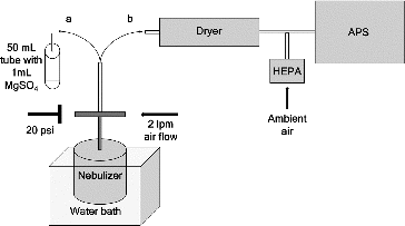
A 1-jet collison nebulizer (BGI, Waltham, MA, USA) was used for the aerosolization experiments. This equipment has been used to aerosolize bacteria previously (Jensen et al. Citation1992; Heidelberg et al. Citation1997; Seaver Citation1999; Brosseau et al. Citation2000; Fergenson et al. Citation2004). Jensen et al. (Citation1992) used a 6-jet version of the collison nebulizer, but the number of jets used in the other publications (Heidelberg et al. Citation1997; Seaver Citation1999; Brosseau et al. Citation2000; Fergenson et al. Citation2004) was not stated. The top of the nebulizer had a tee connection; ambient air flowed into one side of the tee at 2 L per min (lpm), and the other side of the tee was equipped with a female nut and an adjustable plug to obtain a pressure of 20 psi (). This air flow (2 lpm) and pressure setting (20 psi) were selected based on the manufacturer's protocols to minimize damage to the bacteria. An aliquot of 30 mL of bacterial suspension at an OD600 of 0.01 was placed in the nebulizer.
FIG. 1. Schematic of the aerosolization equipment. Aerosol was created in the collison nebulizer with a flow rate of 2 lpm and pressure of 20 psi and collected in 50 mL tubes containing 1 mL of 10 mM MgSO4 for the indirect method (a). For the direct method (b), the aerosol passed through a diffusion dryer and into the Aerodynamic Particle Sizer (APS). Free air was allowed to enter the system through a HEPA filter prior to entering the APS.

Direct Method to Assess the Number of Particles in Aerosols Collected at Four Different Temperatures
The direct method was based on particle counting of aerosols collected at four different temperatures (5°, 15°, 20°, and 30°C). For this method, the aerosol passed through a diffusion dryer model (3062, TSI, Inc., Shoreview, MN, USA) prior to entering the particle counter to remove water droplets (which otherwise could be counted as particles). Water was aerosolized with the nebulizer as a negative control. Particles were counted with the Aerodynamic Particle Sizer (APS) (3321, TSI, Inc., Shoreview, MN, USA). Because the flow rate of the APS was greater than that of the nebulizer, particle-free air was introduced as make-up flow to the APS through an uncontrolled (open) tee connection to the ambient air with a HEPA filter (1602051, TSI, Inc., Shoreview, MN, USA) ().
Indirect Culture Method to Assess the Number of Colony Forming Units (Viable Cells) from Aerosols Collected at Four Different Temperatures
The indirect method was based on plating of aerosols collected at four different temperatures (5°, 15°, 20°, and 30°C). In this method, the aerosol was collected into 1 mL of 10 mM MgSO4 in a sterile 50 mL tube at ambient room temperature by bubbling the aerosol into the liquid in the tube. Each collection was plated on KBC media. Three plates were used for each collection. For each collection, 10-fold dilutions were made for plates of aerosol collected at 15°, 20°, and 30°C. Dilutions were not used for the samples collected at 5°C due to lower aerosol production. All of the plates were incubated at ambient room temperature and CFUs were counted after 72 to 96 h. At less than 72 h, the colonies were not large enough to be counted accurately, and at a time longer than 96 h, the colonies were beginning to grow on top of each other making counting distinct CFUs difficult.
Test Schedule
Collection periods for both methods were 3 min, and three replicates were collected sequentially at each temperature (). The nebulizer jar was submerged in a circulating water bath. The jar was submerged for 30 min with the bath set at 5°C, then collection took place for a total of 9 min (). The bath temperature was then changed, and 21 min passed for the temperature of the sample to stabilize before the next collection period (). The jar remained submerged for the entire experiment. It took approximately 3 min for the water bath to change temperature allowing approximately 18 min for the aerosol to stabilize at each temperature. Temperatures of 5°, 15°, 20°, and 30°C were used. Both strains were tested in alternating order each day. After data were collected for the first strain, the bath temperature was set back to 5°C to begin testing the second strain. It took about 45 min for the bath temperature to return to 5°C. Each sample was in the nebulizer for approximately 2 h. The test schedule was developed based on a series of preliminary laboratory experiments designed to determine (1) the amount of time needed for a sample to reach equilibrium following changing temperature regimes and (2) that fluctuations in aerosolization rates did not vary over the total sampling time.
FIG. 2. Aerosolization test schedule for each strain showing temperature of the water bath (a), testing activity (b), with wait periods in light gray and collection periods in dark gray, and a timeline (c) showing cumulative time. Total test time was 129 min for each strain (ice− 642 and ice+ 892) with an additional 45 min between the first and second strain for the temperature to return to 5°C. A = Temperature, B = activity, C = timeline (not to scale), Teq = temperature equilibrium, and S = 3, 3 min samples (9 min).

Test to Monitor Potential Changes in the CFUs of the Suspensions
To test if the CFUs changed from the beginning to the end of the experiment, the ice− 642 and ice+ 892 strains of P. syringae were prepared at concentrations of 0.01 OD in sterile nanopure water or 10 mM MgSO4. A 30 mL sample was placed in the nebulizer, and aliquots of 10 μL were removed at 0, 3, 6, 9, and 12 h, and were plated on KBC. CFUs were counted 3 days later. The rate of change for concentration of bacteria in the suspension was calculated by subtracting the final concentration of CFUs from the initial concentration divided by the time.
Statistical Analyses
Statistical analyses were conducted with JMP software. ANOVA and Tukey's post-hoc comparison (P < 0.05) were used to test for significant differences between aerosolized samples at different temperatures.
RESULTS
Ice Nucleation Assays to Confirm Ice+ and Ice− Phenotypes
Strain 892 exhibited the ice+ phenotype at all temperatures of incubation, and strain 642 exhibited the ice− phenotype at all temperatures of incubation. Four 12 μL droplets were used for each temperature of incubation (ambient room temperature (∼25°C), 5°, 15°, 20°, and 30°C). For strain 892, the droplets incubated at 20°C froze at −6°C, and the droplets incubated at ambient room temp, 5°, and 15°C froze between −7° and −8°C. The 30°C incubated droplets did not freeze until the temperature reached −10°C. None of the drops of strain 642 froze at any of the temperatures studied.
Direct Method to Assess the Number of Particles in the Aerosol
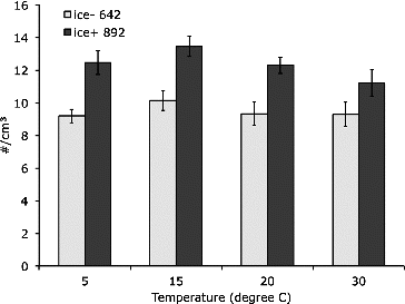
The number of aerosolized particles was not influenced by temperature between 5° and 30°C (). There was no significant difference between any of the temperatures for either strain. In general, the ice+ strain produced a higher number of particles than the ice− strain at each temperature (). There was a significant difference between the strains at 5°, 15°, and 20°C (P < 0.05), but not at 30°C (P = 0.11).
FIG. 3. Concentration of Pseudomonas syringae aerosols produced by a collison nebulizer measured for a 3 min period with an aerodynamic particle sizer at four temperatures for ice− ̶ 642 and ice+ 892 strains with n = 9 in the direct particle counting method. There was a significant difference between the strains at 5°, 15°, and 20°C (P < 0.05), but not at 30°C (P = 0.11).

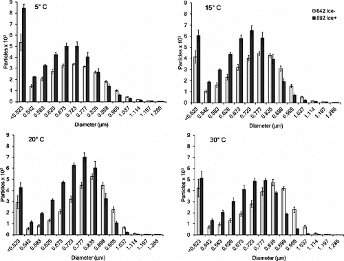
The mean diameter reported by the APS ranged from 0.71 to 0.84 μm, depending on the day of testing (). Cells of P. syringae are rod shaped with a length of 1–5 μm and a width of 0.5–1.5 μm, so submicron aerodynamic diameters are reasonable, especially since the rods are likely to be oriented with their long axis parallel to the direction of flow and they may have been subject to shrinkage in the dryer. Nanopure water was aerosolized as a negative control and the particle counts were very low (around 1 particle per cm3), indicating the dryer was removing a sufficient amount of water. The size distribution of particles differed slightly between the two strains of bacteria (). While more particles were generated by the ice+ strain than by the ice− strain at aerodynamic diameters of 0.8 μm and less, the opposite was generally true for particles >0.8 μm (). There was a pronounced rightward shift of the size distribution, especially at the higher temperatures ().
Indirect Method to Assess the Number of Colony Forming Units (Culturable Cells) from the Aerosol
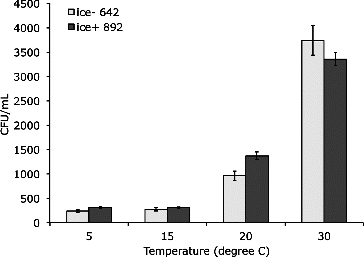
The number of CFUs obtained from aerosolized P. syringae increased with temperature between 5° and 30°C (). In the ice− strain, the temperature pairs 5° and 15°C, 15° and 20°C, and 5° and 20°C were not significantly different (P = 0.472, P = 0.943, and P = 0.189, respectively). The temperature of 30°C was significantly different from all of the other temperatures studied (P < 0.001). For the ice+ strain, the temperature pair of 5° and 15°C was not significantly different (P = 0.999), while all other pairs were significantly different (P < 0.001). Comparing the two strains, a higher number of CFUs were obtained from the aerosolized ice+ strain at all temperatures, with the exception of 30°C (). There was a significant difference in CFUs between the strains for aerosolization at 20°C (P = 0.036). The trend of increasing CFUs with temperature fits an exponential curve with an r-squared value of 0.878 and 0.823 for the ice− and ice+ strains, respectively.
FIG. 5. Indirect method to assess the number of colony forming units (culturable cells) from the aerosol. Two strains were collected in 1 mL of 10 mM MgSO4 over 3 min. Aerosols were produced by a collison nebulizer at four temperatures for ice− 642 and ice+ 892 strains with n = 9, except ice̶ at 15°C where n = 6.There was a significant difference between the culturability of the aerosols between the strains at 20°C (P = 0.036).

Confirming CFUs of Suspensions in the Nebulizer Over Test Duration
The concentrations of the bacterial suspensions in MgSO4 were higher than the starting concentration at 3, 6, and 12 h, but the concentration of bacteria did not increase consistently with time in the nebulizer (e.g., concentrations of bacteria were higher for the strains at 6 h than at 12 h) (as in the online supplementary Figure S1). In general, concentrations decreased for the suspensions in H2O over all times tested (Figure S1). The volumes of the suspensions in the nebulizer were measured in a graduated cylinder before and after the 2 h aerosolization period, and the amount lost was less than the resolution of the graduated cylinder (data not shown). After 12 h of aerosolization in the nebulizer, the volume of the suspension decreased from 30 mL to ∼20 mL.
DISCUSSION
Freshwater aerosols are understudied, and few efforts have tracked the life history of microorganisms in aerosols. Here, we show that (1) the culturability (viability) of two strains of P. syringae aerosolized in a collison nebulizer is impacted by temperature and (2) particle counts of the aerosols are not impacted by temperature. Our experiments were designed to test the hypotheses that: (1) the production of aerosolized P. syringae particles will increase with temperature, (2) the number of viable aerosolized P. syringae will increase with temperature, and (3) an ice-nucleating strain (ice+) of P. syringae will aerosolize at a greater rate than a nonice nucleating strain (ice−). Results from our experiments suggest that P. syringae has the potential to be aerosolized from natural aquatic environments (streams, rivers, ponds, and lakes—known reservoirs of P. syringae (Morris et al. Citation2008)) at a variety of temperatures. This process may be an important part of P. syringae's life cycle. Strains of P. syringae have been found in the atmosphere and in aquatic environments (Morris et al. Citation2013), yet the movement between these two environments and factors affecting the flux has not been studied in detail. Aerosolized bacteria are known to be transported in the atmosphere (Franc Citation1988; Morris et al. Citation2013). The results described here improve our understanding of the aerosolization of P. syringae from aquatic environments, and could be applied to a generalized model of bacterial aerosolization flux in the future.
Aerosolization of P. syringae was not influenced by water temperatures between 5° and 30°C. Previous studies have suggested that temperature may increase bacterial aerosol concentration above plant canopies (Lighthart and Shaffer Citation1994; Lindemann and Upper Citation1985). Thus, our observation that aerosolization was not influenced by temperature was unexpected. Our work suggests that aerosolization of P. syringae in natural aqueous environments would not change with seasonal temperature changes. Unknown, however, are the contributions of other factors correlated with seasonal changes, such as changes in water velocity and turbulence (Sharma et al. Citation2007), that may affect aerosolization during different times of the year. Additionally, the temperature at the surface of the water in natural environments may be higher because of direct solar radiation.
In general, the culturability of P. syringae in aerosols increased with temperature between 5 and 30°C. This observation is supported by a shorter doubling time for P. syringae at higher temperatures (between 0 and 36°C). Previous work has shown that the doubling rate of P. syringae is around 10.45 h at 5.3°C, and 1.3 h at 26.0°C (Young et al. Citation1977). In natural aqueous systems, viable aerosolized particles of P. syringae might be expected to increase with temperature and may therefore change seasonally. Viable cells of P. syringae are essential for infection and reproductive success, but both viable and nonviable bacteria can nucleate ice. Aerosolized cells of P. syringae have been shown to lose viability after UV-A exposure, but retain their ice nucleation ability (Attard et al. Citation2012).
The ice+ strain produced greater total numbers of particles than did the ice– strain at all temperatures studied (5° to 30°C). This observation suggests a possible connection between ice nucleation phenotype and aerosol production. Provided the same volume of liquid is aerosolized, the same total number of bacteria must be aerosolized in either case, but the strain affects the resulting size of the aerosols. Perhaps the bacteria of the ice− strain stick together more, such that two or more cells comprise a single aerosol, while the bacteria of the ice+ strain tend toward single-cell aerosols. For some bacteria, an aggregation protein has been shown to influence the degree of aggregation, and the expression of different surface antigens appears to change hydrophobicity (Lindahl et al. Citation1981; Wells et al. Citation2000). In this scenario, the ice− strain would produce a smaller total number of aerosols and have a rightward shifted size distribution compared to the ice+ strain. It is also possible that there are changes in the relative solubility of each of the strains that could be related to phenotype differences, but this was not examined in this study.
At 30°C, the ice+ 892 strain did not freeze until it reached −10°C, which was 2 to 4°C cooler than bacteria incubated at the other temperatures. In the direct particle counting method, 30°C was the only temperature which did not report a significant difference between strains, while in the indirect culture method 30°C was the only temperature which did not show higher aerosol production for the ice+ strain. Perhaps the ice nucleation activity is reduced at 30°C, and this is reflected in aerosol production. It is possible that the ice+ strain may be suited for transport and survival in the atmosphere. Harsh atmospheric conditions including lack of water are harmful to bacteria, especially nonspore forming bacteria such as P. syringae (Chi and Li Citation2007; Polymenakou Citation2012). Morris et al. (Citation2011) found all strains of P. syringae collected from snow and rain were ice nucleation active, but not all strains collected from other sources including plants and water were ice nucleation active (Morris et al. Citation2008; Morris et al. Citation2011). Monteil et al. (Citation2014) found all strains collected from snow were ice nucleation active while some strains collected from rain were not ice nucleation active. This may indicate that the ice nucleation phenotype favors survival in atmospheric conditions either by preferentially allowing the bacteria to enter the atmosphere or increasing survival once bacteria have entered the atmosphere. Even if the difference between the two tested strains is not due to the IN phenotype, our data indicate that aerosolization rates may not be constant for all strains of the same species. Both of the strains used in this study are closely related and part of the 2c phylogenic subgroup (Clarke et al. Citation2010). The difference in aerosol production between these two strains, regardless of the connection to IN phenotype, suggests that different strains are aerosolized at different rates in natural environments. Thus, future models of the aerosolization of P. syringae from natural aquatic environments would need to account for this variability. It should be noted that bacterial suspensions were plated before and after the aerosolization experiments, and there was no significant increase in concentration for the ice+ 892 strain compared to the ice− 642 strain, indicating differences in the concentration of the suspension were not the cause of differences in aerosol production between the strains. The concentrations of the bacterial suspensions in MgSO4 were higher than the starting concentration at 3, 6, and 12 h, but the concentration of bacteria did not increase consistently with time in the nebulizer (e.g., concentrations of bacteria were higher for the strains at 6 h than at 12 h), and concentrations generally decreased for the suspensions in H2O over all times tested. Thus, changes in aerosol production are not likely a result of changes in bacterial concentration in the suspension. Use of other nebulizers may increase the robustness of our results but that was beyond the scope of this study. A number of studies have generated bacteria aerosols using only a collison nebulizer (Heidelberg et al. Citation1997; Brosseau et al. Citation2000; Fergenson et al. Citation2004).
Future work should include knocking out the IN gene in the ice+ 892 strain, and performing aerosolization experiments comparing the ice+ strain with the genetically modified ice- strain. Since the only difference between the two strains will be the INA gene, differences between strains can then be interpreted as being directly caused by the INA gene. Other strains of P. syringae with ice− and ice+ phenotypes could also be subjected to similar aerosolization experiments. Future work aims to examine the aerosolization of P. syringae from aqueous field environments. Such studies may assist in elucidating the mechanisms of natural aerosol production in the future, a necessary component of a comprehensive understanding of the aerosolization process and the ecology of microbial life in the atmosphere.
SUPPLEMENTAL MATERIAL
Supplemental data for this article can be accessed on the publisher's website.
UAST_1010636_Supplemental_Information.zip
Download Zip (75.2 KB)ACKNOWLEDGMENTS
The authors thank A. Tiwari, K. Failor, and C. Clarke for their technical expertise. Any opinions, findings, and conclusions or recommendations expressed in this material are those of the authors and do not necessarily reflect the views of the National Science Foundation.
Funding
This research was supported in part by the National Science Foundation (NSF) under Grant Nos. DEB-1241068 (Dimensions: Collaborative Research: Research on Airborne Ice-Nucleating Species [RAINS]) and DGE-0966125 (IGERT: MultiScale Transport in Environmental and Physiological Systems [MultiSTEPS]).
REFERENCES
- Aller, J. Y., Kuznetsova, M. R., Jahns, C. J., and Kemp, P. F. (2005). The Sea Surface Microlayer as a Source of Viral and Bacterial Enrichment in Marine Aerosols. J. Aerosol Sci., 36:801–812.
- Amato, P., Parazols, M., Sancelme, M., Laj, P., Mailhot, G., and Delort, A. M. (2007). Microorganisms Isolated from the Water Phase of Tropospheric Clouds at the Puy de Dôme: Major Groups and Growth Abilities at Low Temperatures. FEMS Microbiol. Ecol., 59:242–254.
- Attard, E., Yang, H., Delort, A.-M., Amato, P., Pöschl, U., Glaux, C., Koop, T., and Morris, C. (2012). Effects of Atmospheric Conditions on Ice Nucleation Activity of Pseudomonas. Atmos. Chem. Phys., 12:10667–10677.
- Baylor, E., Baylor, M., Blanchard, D. C., Syzdek, L. D., and Appel, C. (1977). Virus Transfer from Surf to Wind. Science, 198:575–580.
- Bigg, E., and Miles, G. (1964). The Results of Large-Scale Measurements of Natural Ice Nuclei. J. Atmos. Sci., 21:396–403.
- Bigg, E. K., (2007). Sources, Nature and Influence on Climate of Marine Airborne Particles. Environ. Chem., 4:155–161.
- Blanchard, D. C., (1989). The Ejection of Drops From the Sea and Their Enrichment With Bacteria and Other Materials: A Review. Estuaries, 12:127–137.
- Blot, R., Clarke, A., Freitag, S., Kapustin, V., Howell, S., Jensen, J., Shank, L., McNaughton, C., and Brekhovskikh, V. (2013). Ultrafine Sea Spray Aerosol Over the South Sastern Pacific: Open-Ocean Contributions to Marine Boundary Layer CCN. Sea, 13:3279–3322.
- Brosseau, L. M., Vesley, D., Rice, N., Goodell, K., Nellis, M., and Hairston, P. (2000). Differences in Detected Fluorescence Among Several Bacterial Species Measured with a Direct-Reading Particle Sizer and Fluorescence Detector. Aerosol Sci. Technol., 32:545–558.
- Cavalli, F., Facchini, M., Decesari, S., Mircea, M., Emblico, L., Fuzzi, S., Ceburnis, D., Yoon, Y., O’Dowd, C., and Putaud, J. P. (2004). Advances in Characterization of Size-Resolved Organic Matter in Marine Aerosol Over the North Atlantic. J. Geophys. Res.: Atmos. (1984–2012) 109(D24, 27).
- Chi, M.-C., Li, C.-S., (2007). Fluorochrome in Monitoring Atmospheric Bioaerosols and Correlations With Meteorological Factors and Air Pollutants. Aerosol Sci. Technol., 41:672–678.
- Claeys, M., Wang, W., Vermeylen, R., Kourtchev, I., Chi, X., Farhat, Y., Surratt, J. D., Gómez-González, Y., Sciare, J., and Maenhaut, W. (2010). Chemical Characterisation of Marine Aerosol at Amsterdam Island During the Austral Summer of 2006–2007. J. Aerosol Sci., 41:13–22.
- Clarke, C. R., Cai, R., Studholme, D. J., Guttman, D. S., and Vinatzer, B. A. (2010). Pseudomonas syringae Strains Naturally Lacking the Classical P. syringae hrp/hrc Locus are Common Leaf Colonizers Equipped With an Atypical Type III Secretion System. Mol. Plant–Microbe Interact., 23:198–210.
- Cochet, N., and Widehem, P. (2000). Ice Crystallization by Pseudomonas syringae. Appl. Microbiol. BioTechnol., 54:153–161.
- Constantinidou, H., Hirano, S., Baker, L., and Upper, C. (1990). Atmospheric Dispersal of Ice Ucleation-Active Bacteria: The Role of Rain. Phytopathology, 80:934–937.
- DeMott, P. J., Sassen, K., Poellot, M. R., Baumgardner, D., Rogers, D. C., Brooks, S. D., Prenni, A. J., and Kreidenweis, S. M. (2003). African Dust Aerosols as Atmospheric Ice Nuclei. Geophys. Res. Lett. 30.
- Després, V. R., Huffman, J. A., Burrows, S. M., Hoose, C., Safatov, A. S., Buryak, G., Fröhlich-Nowoisky, J., Elbert, W., Andreae, M. O., and Pöschl, U. (2012). Primary Biological Aerosol Particles in the Atmosphere: A Review. Tellus B 64.
- Diehl, K., Matthias-Maser, S., Jaenicke, R., and Mitra, S. (2002). The Ice Nucleating Ability of Pollen: Part II. Laboratory Studies in Immersion and Contact Freezing Modes. Atmos. Res., 61:125–133.
- Edwards, G., and Evans, L. (1960). Ice Nucleation by Silver Iodide: I. Freezing vs Sublimation. J. Meteorol., 17:627–634.
- Fahlgren, C., Bratbak, G., Sandaa, R.-A., Thyrhaug, R., and Zweifel, U. L. (2011). Diversity of Airborne Bacteria in Samples Collected Using Different Devices for Aerosol Collection. Aerobiologia, 27:107–120.
- Fergenson, D. P., Pitesky, M. E., Tobias, H. J., Steele, P. T., Czerwieniec, G. A., Russell, S. C., Lebrilla, C. B., Horn, J. M., Coffee, K. R., and Srivastava, A. (2004). Reagentless Detection and Classification of Individual Bioaerosol Particles in Seconds. Anal. Chem., 76:373–378.
- Franc, G. D., (1988). Long Distance Transport of Erwinia Carotovora in the Atmosphere and Surface Water. Colorado State University.
- Fu, P., Kawamura, K., and Miura, K. (2011). Molecular Characterization of Marine Organic Aerosols Collected During A Round-the-World Cruise. J. Geophys. Res.: Atmos. (1984–2012) 116. DOI:10.1029/2011JD015604.
- Heidelberg, J., Shahamat, M., Levin, M., Rahman, I., Stelma, G., Grim, C., and Colwell, R., (1997). Effect of Aerosolization on Culturability and Viability of Gram-Negative Bacteria. Appl. Environ. Microbiol., 63:3585–3588.
- Hirano, S. S., and Upper, C. D. (2000). Bacteria in the Leaf Ecosystem with Emphasis on Pseudomonas syringae—a Pathogen, Ice Nucleus, and Epiphyte. Microbiol. Mol. Biol. Rev., 64:624–653.
- Hoose, C., Kristjánsson, J., and Burrows, S. (2010). How Important is Biological Ice Nucleation in Clouds on a Global Scale? Environ. Res. Lett., 5:024009.
- Howell, W. E., (1949). The Growth of Cloud Drops in Uniformly Cooled Air. J. Meteorol., 6:134–149.
- Hudson, J. G., (1993). Cloud Condensation Nuclei. J. Appl. Meteorol., 32. DOI:10.1029/GL009i001p00094.
- Jayaweera, K., and Flanagan, P. (1982). Investigations on Biogenic Ice Nuclei in the Arctic Atmosphere. Geophys. Res. Lett., 9:94–97.
- Jensen, P. A., Todd, W. F., Davis, G. N., and Scarpino, P. V. (1992). Evaluation of Eight Bioaerosol Samplers Challenged With Aerosols of Free Bacteria. Am. Ind. Hyg. Assoc. J., 53:660–667.
- Kozloff, L., Schofield, M., and Lute, M. (1983). Ice Nucleating Activity of Pseudomonas Syringae and Erwinia Herbicola. J. Bacteriol., 153:222–231.
- Kozloff, L., Turner, M., and Arellano, F. (1991). Formation of Bacterial Membrane Ice-Nucleating Lipoglycoprotein Complexes. J. Bacteriol., 173:6528–6536.
- Lighthart, B., (1997). The Ecology of Bacteria in the Alfresco Atmosphere. FEMS Microbiol. Ecol., 23:263–274.
- Lighthart, B., and Shaffer, B. (1994). Bacterial Flux from Chaparral Into the Atmosphere in Mid-Summer at a High Desert Location. Atmos. Environ., 28:1267–1274.
- Lindahl, M., Faris, A., Wadstr m, T., and Hjertén, S. (1981). A New Test Based on ‘Satling Out’ to Measure Relative Surface Hydrophobicity of Bacterial Cells. Biochim. Biophys. Acta, 677:471–476.
- Lindemann, J., Constantinidou, H. A., Barchet, W. R., and Upper, C. D. (1982). Plants as Sources of Airborne Bacteria, Including Ice Nucleation-Active Bacteria. Appl. Environ. Microbiol., 44:1059–1063.
- Lindemann, J., and Upper, C. (1985). Aerial Dispersal of Epiphytic Bacteria Over Bean Plants. Appl. Environ. Microbiol., 50:1229–1232.
- Lindow, S., (1983). The Role of Bacterial Ice Nucleation in Frost Injury to Plants. Ann. Rev. Phytopathol., 21:363–384.
- Lindow, S., Arny, D., and Upper, C. (1978). Erwinia Herbicola: A Bacterial Ice Nucleus Active in Increasing Frost Injury to Corn. Phytopathology, 68:523–527.
- Lohmann, U., and Diehl, K. (2006). Sensitivity Studies of the Importance of Dust Ice Nuclei for the Indirect Aerosol Effect on Stratiform Mixed-Phase Clouds. J. Atmos. Sci., 63:968–982.
- Lundheim, R., (2002). Physiological and Ecological Significance of Biological Ice Nucleators. Philos. Trans. R. Soc. London. Ser. B: Biol. Sci., 357:937–943.
- Maki, L. R., Galyan, E. L., Chang-Chien, M.-M., and Caldwell, D. R. (1974). Ice Nucleation Induced by Pseudomonas syringae. Appl. Microbiol., 28:456–459.
- Marks, R., Kruczalak, K., Jankowska, K., and Michalska, M. (2001). Bacteria and Fungi in Air Over the Gulf of Gdańsk and Baltic Sea. J. Aerosol Sci., 32:237–250.
- Mason, B., (1952). The Spontaneous Crystallization of Supercooled Water. Quart. J. R. Meteorol. Soc., 78:22–27.
- Mason, B., and Ludlam, F. (1951). The Microphysics of Clouds. Rep. Progr. Phys., 14:147–195.
- Mohan, S., and Schaad, N. (1987). An Improved Agar Plating Assay for Detecting Pseudomonas syringae pv. syringae and P. s. pv. phaseolicola in Contaminated bean Seed. Phytopathology, 77:1390–1395.
- Monteil, C. L., Bardin, M., and Morris, C. E. (2014). Features of Air Masses Associated With the Deposition of Pseudomonas syringae and Botrytis Cinerea by Rain and Snowfall. The ISME J. 8:2290–2304.
- Morris, C., Sands, D., Bardin, M., Jaenicke, R., Vogel, B., Leyronas, C., Ariya, P., and Psenner, R. (2011). Microbiology and Atmospheric Processes: Research Challenges Concerning the Impact of Airborne Micro-Organisms on the Atmosphere and Climate. Biogeosciences, 8:17–25.
- Morris, C. E., Kinkel, L. L., Xiao, K., Prior, P., and Sands, D. C. (2007). Surprising Niche for the Plant Pathogen Pseudomonas syringae. Infect. Genet. Evol., 7:84–92.
- Morris, C. E., Monteil, C. L., and Berge, O. (2013). The Life History of Pseudomonas syringae: Linking Agriculture to Earth System Processes. Ann. Rev. Phytopathol..
- Morris, C. E., Sands, D. C., Vinatzer, B. A., Glaux, C., Guilbaud, C., Buffière, A., Yan, S., Dominguez, H., and Thompson, B. M. (2008). The Life History of the Plant Pathogen Pseudomonas syringae is Linked to the Water Cycle. ISME J., 2:321–334.
- Mossop, S., (1955). The Freezing of Supercooled Water. Proc. Phys. Soc.. B, 68:193.
- Murray, B., O’Sullivan, D., Atkinson, J., and Webb, M. (2012). Ice Nucleation by Particles Immersed in Supercooled Cloud Droplets. Chem. Soc. Rev., 41:6519–6554.
- Polymenakou, P. N., (2012). Atmosphere: A Source of Pathogenic or Beneficial Microbes? Atmosphere, 3:87–102.
- Pósfai, M., Li, J., Anderson, J. R., and Buseck, P. R. (2003). Aerosol Bacteria Over the Southern Ocean During Ace-1. Atmos. Res., 66:231–240.
- Pouleur, S., Richard, C., Martin, J.-G., and Antoun, H. (1992). Ice Nucleation Activity in Fusarium acuminatum and Fusarium avenaceum. Appl. Environ. Microbiol., 58:2960–2964.
- Pratt, K. A., DeMott, P. J., French, J. R., Wang, Z., Westphal, D. L., Heymsfield, A. J., Twohy, C. H., Prenni, A. J., and Prather, K. A. (2009). In Situ Detection of Biological Particles in Cloud Ice-Crystals. Nature Geosci., 2:398–401.
- Riffaud, C. M. H., and Morris, C. E. (2002). Detection of Pseudomonas syringae pv. aptata in Irrigation Water Retention Basins by Immunofluorescence Colony-Staining. Eur. J. Plant Pathol., 108:539–545.
- Rixen, C., Stoeckli, V., and Ammann, W. (2003). Does Artificial Snow Production Affect Soil and Vegetation of Ski Pistes? A Review. Perspect. Plant Ecol. Evol.Syst., 5:219–230.
- Sands, D., Langhans, V., Scharen, A., De Smet, G., (1982). The Association Between Bacteria and Rain and Possible Resultant Meteorological Implications. Idojaras.
- Schaefer, V. J., (1952). Formation of Ice Crystals in Ordinary and Nuclei-Free Air. Ind. Eng. Chem., 44:1300–1304.
- Seaver, M., (1999). Size and Fluorescence Measurements for Field Detection of Biological Aerosols. Aerosol Sci. Technol., 30:174–185.
- Sharma, N. K., Rai, A. K., Singh, S., and Brown, R. M. (2007). Airborne Algae: Their Present Status and Relevance. J. Phycol., 43:615–627.
- Vali, G., Christensen, M., Fresh, R., Galyan, E., Maki, L., and Schnell, R. (1976). Biogenic Ice Nuclei. Part II: Bacterial Sources. J. Atmos. Sci., 33:1565–1570.
- Vonnegut, B., (1947). The Nucleation of Ice Formation by Silver Iodide. J. Appl.Phys., 18:593–595.
- Weidner, T., (2013). Uncovering the Tricks of Nature's Ice-Seeding Bacteria. Catherine Meyers [cited 1 December 2013]. Available from http://www.eurekalert.org/pub_releases/2013-10/aiop-utt102313.php.
- Wells, C.L, Moore, E. A., Hoag, J. A., Hirt, H., Dunny, G. M., and Erlandsen, S. L. (2000). Inducible Expression of Enterococcus faecalis Aggregation Substance Surface Protein Facilitates Bacterial Internalization by Cultured Enterocytes. Infect. Immun., 68:7190–7194.
- Young, J., Luketina, R., and Marshall, A. (1977). The Effects on Temperature on Growth In Vitro of Pseudomonas syringae and Xanthomonas pruni. J. Appl. Bacteriol., 42:345–354.
- Zhao, Y., Aarnink, A. J., Doornenbal, P., Huynh, T. T., Koerkamp, P.W.G., de Jong, M. C., and Landman, W. J. (2011). Investigation of the Efficiencies of Bioaerosol Samplers for Collecting Aerosolized Bacteria Using a Fluorescent Tracer. I: Effects of Non-Sampling Processes on Bacterial Culturability. Aerosol Sci. Technol., 45:423–431.

