Abstract
The Danish Breast Cancer Cooperative Group's register containing data from about 75 000 patients undergoing surgery for primary invasive breast cancer from 1978–2006 has been examined for classical pathological variables. During that period the diagnostic approach of malignant breast tumours experienced a major shift from invasive surgical procedures to non invasive procedures with an increase of the proportion of both fine needle aspiration biopsy and core needle biopsy. According to variation in tumour size there has been a slight increase in tumours <10 mm and a significant decrease in tumours >50mm from 7 to 4%. The distribution of the histological subtypes of malignant breast tumours has been almost unchanged. We found however a significant increase in the number of high grade tumours. A large increase in the number of removed axillary lymph nodes from 1989–2001 is related to improved surgical procedure. The subsequent decline from 2002–2006 is the result of the introduction of the sentinel node technique. In the five-year period 2002–2006 following the introduction of sentinel node technique, the frequency of patients having at least one lymph node metastases (48.7%) was higher than in the preceding five-year period 1997–2001 (44.4%, p=0.0087).
The Danish Breast Cancer Cooperative Group (DBCG) was organized in 1977 with the aim to provide guidelines for treatment of patients with primary breast cancer, to conduct nationwide clinical trials of therapy of primary breast cancer and to evaluate treatment programmes Citation[1]. A population based clinical database was founded and diagnostic, therapeutic and follow up data has since then been collected by use of standardized forms. The DBCG register includes close to 100% of all patients surgically treated by lumpectomy or mastectomy in Denmark. The purpose of the present work is to investigate the variation of different classical pathological variables from 1978–2006.
Material and methods
This nationwide investigation is based on pathological data from 75 107 patients surgically treated for primary invasive breast cancer by lumpectomy or mastectomy from 1978–2006 and entered the DBCG register. During this period breast pathology was performed at 30 pathological departments (including 3 private laboratories). All of these were active from 1978/79 except one department that came into service in 1986 (Roskilde). Fourteen departments ceased activity before 2006.
Patients were initially diagnosed by either surgical biopsy or needle biopsy. Patients′ tumour characteristics include histological subtype, malignancy grade, tumour size, lymph node status, vascular invasion and the registration of more than one tumour focus in the breast, if present.
Histological subtype based on the WHO-classification Citation[2–4] is described as invasive ductal carcinoma, invasive lobular carcinoma, and “invasive other”. The group “invasive other” includes mucinous, medullary, papillary, tubular, adenoid cystic, secretory, apocrine, metaplastic, and other special types not mentioned in the WHO classification.
In Denmark the entity invasive ductal carcinoma (not otherwise specified) has traditionally been graded in three histological subgroups; grade I to III using a grading system close to and, from 1989, almost identical to the grading system described by Elston and Ellis Citation[5].
The tumour size was based on the surgeon's or the pathologist's measurement until 1982. After 1982, the tumour size was estimated by the pathologist only as a combined macroscopic and microscopic measurement.
The presence of more than one tumour in the breast has been recorded differently over time. Multicentric carcinoma (MC) defined as the presence of tumour in at least two different quadrants of the breast was recorded from 1982–1989. There was no record of MC from 1990–1995. From 1996 until December 2001 multifocal carcinoma (MF), defined as more than one tumour focus separated from the index tumour by benign mammary tissue was recorded. There was no demand of any minimum distance between the individual foci in this period. Since December 2001, MF has been defined as the localisation of more than one tumour separated by benign mammary tissue with a distance of >20 mm, whereas invasive carcinoma separated by benign mammary tissue with a distance of ≤20 mm was defined as unifocal carcinoma with satellite.
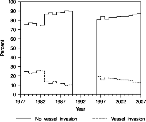
The presence of vascular invasion was recorded in the period 1977–1989 and again from 1996 onwards. During the period 1977–1982, there was no specified definition on vascular invasion. From 1982–1989, definitive vascular invasion was defined as tumour cells in vascular lumen with coexistence of fibrintrombus and/or endothelial proliferation and/or fibrosis. Questionable vascular invasion was defined as presence of tumour cells in vascular lumen without any of the above-mentioned reactions.
From 1996 vascular invasion was defined as the presence of tumour cells localised within endothelial lined cavities beyond the periphery of the invasive carcinoma.
The number of axillary lymph nodes entered DBCG includes both sentinel lymph node (SN) as well as the total number of lymph nodes (LN) after complete axillary lymph node dissection (ALDN).
In 1989 DBCG implemented recommendations on ALDN to include at least 10 LN in order to improve staging of the disease with curative and diagnostic purposes. The SN technique was introduced as an experimental method in 1998 using patent blue dye and/or radioactive tracer, and the method was implemented nationwide between November 2001 and October 2004.
The 2002 edition of the TNM classification uses 2.0 mm as the cut-off between micro- and macrometastases Citation[6]. Micrometastases are defined as clusters of more than 10 tumour cells or between 0.2 mm and ≤2 mm.
Isolated tumour cells are defined as single tumour cells and/or small clusters of tumour cells with a maximum count of 10 tumour cells. Both the identification of micrometastases and/or isolated tumour cells in the SN results in complete ALDN.
The vast majority of breast cancer cases were usually clinically detected, but from 1991 and 1993, respectively, screening by mammography was offered to women aged 50–69 years in two regions, the municipality of Copenhagen and the county of Funen. The population of these two regions accounts for approximately 20% of the total Danish population.
Statistics
For each parameter the distribution by year was determined and the results were presented as plots of percentage by year. Investigations of time trends were made for the parameters: tumour size, grade, and positive lymph nodes. Observations were transformed into indicators of tumour size >20 mm, tumour size >50 mm, histological grade II and III, histological grade III, and the number of positive lymph nodes ≥1 (N + ) and were grouped by pathological department and year of examination. Regression analyses of indicator value by year were done by mixed effects logistic models. Analyses were made for a subgroup of 18 departments, which were in service during the entire period 1978–2006 except for one department that was initiated in 1979 and another three departments that ceased activity in 2005 or 2006. This subgroup included 63 818 patients. The analysis of tumour size was done for the period 1983–2006 and the analysis of N+ were done for the period 1978–2001. For N+, an additional analysis was done for comparison of the periods 1997–2001 and 2002–2006, i.e. before and after the implementation of sentinel node technique. The analysis of grade II and III was done for the period 1990–2006 where histological grade has been a criterion in the classification of high-risk patients eligible for adjuvant therapy. Models were fitted to data and maximum likelihood estimates of parameters were obtained by the Proc Nlmixed of SAS ver. 8.2 (SAS Institute, Cary, NC, USA).
Results
Overall there has been an increase of the total number of diagnosed breast cancer patients in Denmark from 1 462 cases in 1978 increasing to a peak level in 1992 with 2 948 cases. After a short decline, the rate rose steadily to another peak level in 2002 with 3 562 DBCG registered cases. Since then the incidence rate has stabilized through 2002–2004 and is presently slightly declining to 3 340 cases in 2006.
Since 1982 the type of biopsy has been specified (n = 59528). Excision biopsy was the diagnostic method of choice in the eighties and the first half of the nineties, but declined from 75% to only 5% in 2006. The proportions of core needle biopsy (CNB) and fine needle aspiration biopsy (FNAB) have increased from 10% in 1983 to 25% in 1996. Since then the proportion of FNAB has been fairly constant (25 to 30%), while the proportion of CNB presently dominates the triple diagnostic approach by 65%.
The surgical procedure consists of either mastectomy or lumpectomy (n = 75 107). The use of mastectomy has declined from almost 100% in 1978 to 50% in 2006 while the proportion of lumpectomy has increased from nearly zero in 1978 to 50% in 2006.
The distribution of tumour size is presented in (n = 71 698). Regarding tumours ≤10 mm and >50 mm, they each constituted 10% from 1978 to 1982. Since 1983 there has been a slight increase in the percentage of patients with tumour size ≤10 mm (from 12 to 14%) and a slight decrease in tumour size >50 mm (from 7 to 4%). The analysis of time trends for tumour size >50 mm showed that the effect of examination year was highly significant (p=0.0001, ). The estimated slope by year had odds ratio of 0.971 (95% CI: 0.959-0.984), which showed that the frequency of large tumours decreased relatively by 3% per year. When comparing the frequencies of tumour size >20 mm versus ≤20 mm the time trend was not significant (P=0.22). From 1978 to 1982 the percentage of patients with tumours ranging from 21–50 mm was almost 50% and the percentage of the patients with tumours between 11–20 mm was approximately 28%. Since 1982 the proportional occurrence of both of the last mentioned tumour sizes has remained stable at about 40%.
Figure 1. Tumour size. The percentage of patients with tumour size <10 mm, 11-20 mm, 21-50 mm, and >50 mm in diameter (n = 71 698).
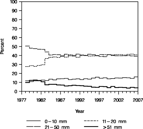
Table I. Analyses of time trends of tumor size, histological grade, and lymph node metastatases by mixed effects logistic regression models.
The frequency of MC and/or MF was 16.3% in the period 1982–1989, 14.1% from 1996–2001, and 9.8% in 2002–2006 among a total of 49 861 patients. The frequency of unifocal carcinomas with satellites was 12.7% in 2002–2006 (n = 16 088).
The percentage of the patients with invasive ductal carcinoma has been fairly constant since 1983 constituting approximately 82% of all histological subtypes (n = 75 107). The proportion of invasive lobular carcinoma has increased from 3–4% in 1978 to 11% in 1989 and has since then remained fairly stable. The group “invasive other” constitutes 7% of the histological subtypes.
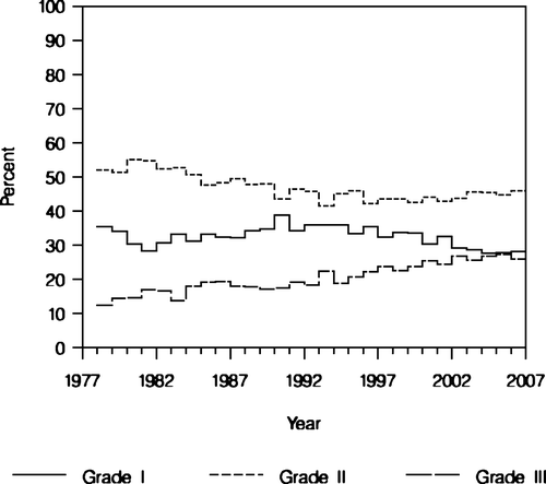
Regarding histological grade, the proportion of grade I (well-differentiated) tumours accounts for 30% while the proportion of grade II (intermediate-differentiated) tumours has decreased slightly from 55 to 45%. Finally, grade III (low-differentiated) tumours constituted an increasing proportion of the population 15 to 25% (n = 59 722) (). When analysing the single components contributing to the grading system it appears that the proportion of tumours with major nuclear polymorphism increased significantly during time. Likewise an increase in number of mitosis was observed while the distribution of tubular growth pattern remained stable. Since 1989 premenopausal patients and since 1999 all patients with tumours otherwise defined as low risk (node negative, tumour size ≤20 mm) were allocated to the high risk group eligible for adjuvant therapy Citation[7], if the tumour was classified as grade II or III. Since 1989, the distribution of grade I as opposed to the combination of grade II and III remained constant (p = 0.16, ). With regard to grade III tumours, the proportion increased significantly during the period 1978–2006 (p = 0.012). The estimated slope by year had odds ratio of 1.030 (95% CI: 1.008–1.053), which showed that frequency of high grade tumours increased relatively by 3% per year.
Figure 2. Histological grade of invasive ductal carcinoma (NOS). The percentage of patients with malignancy grade I, grade II, and grade III (n = 59 722).
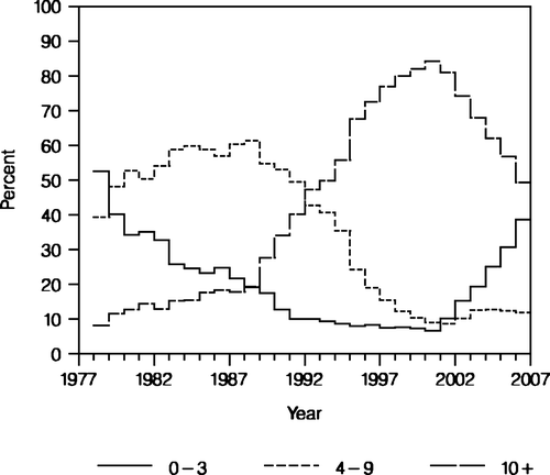
The number of dissected axillary LN has changed dramatically. As shown in , since 1989 the proportion of patients with ALND comprising at least 10 LN increased until 2001 (n = 59 722). Since the introduction of the SN technique the number of dissected LN has decreased markedly. For SN positive patients who all underwent ALDN, the proportion of 10 or more examined LN is approximately 80% (data not shown).
Figure 3. Number of removed axillary lymph nodes. Percentage of patients with 0-3 lymph nodes, 4-9 lymph nodes, and 10 or more lymph nodes (n = 74 957).
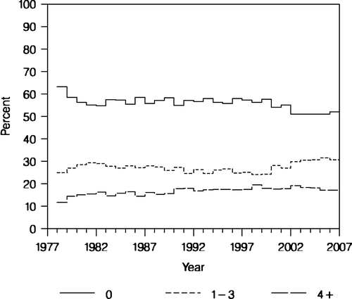
The distribution of node negative patients, node positive patients with 1–3 positive nodes and node positive patients having ≥4 positive nodes is shown in (n = 74 961). There has been a slight decline in the number of LN negative patients and a slight increase in the percentage of LN positive patients (1–3 positive nodes). In the analysis of time trends for patients having at least one lymph node metastasis diagnosed between 1978 and 2001, the effect of examination year was not significant (p = 0.69, ). The additional analysis that compared the 5-year periods before and after the implementation of the sentinel node technique showed that the percentage of patients having at least one lymph node metastasis increased significantly from 44.4% (95% CI: 42.7–46.0) in 1997–2001 to 48.7% (95% CI: 46.4–51.0) in 2002–2006 (p = 0.00087).
Figure 4. Number of tumour positive lymph nodes. Percentage of node negative patients, patients with 1-3 positive nodes, and patients with 4 or more positive nodes (n = 74 961).
Since 1989 the incidence of micrometastases has been recorded. Until 2001 micrometastases was found in 5.1% of the patients. From 2002–2006, the frequency increased to 8.4%. From January 2004, isolated tumour cells were recorded solely as isolated tumour cells and the corresponding involved lymph node as node negative. This phenomenon was recorded in 3.6% of the patients.
The registration of vascular invasion is demonstrated in (n = 57 090). From 1977–1989 the definition of vascular invasion was interchangeable and consequently the registered rates are partly unreliable. From 1996 the registration of peritumoral vascular invasion (PVI) was definite and the rate of PVI is slightly declining from approximately 20% in 1996 to 13% in 2006.
Discussion
In general breast cancer incidence rate in Denmark has risen since 1977. The peak level observed in the Danish population in the first half of the 1990s is consistent with the adoption of screening mammography in the two regions of Denmark. The second peak in 2002 may partly correspond to increases in menopausal hormone therapy throughout the 1990s Citation[8].
The diagnostic procedure of breast tumours has chanced markedly during the years. The establishment of non-operative diagnosis achieved by means of the triple approach combining the results of imaging and clinical examination with either CNB or FNAB has been implemented as clinical routine procedure hence the increased proportion of registered FNAC and CNB. The advantage of CNB as compared to FNAC comprises the ability of performing immunohistochemical analysis for hormone-receptors and HER2.
The change in measurement of tumour size from surgeon or pathologist to pathologist only in 1983 might explain the sudden change in the proportions between the two tumour dimensions 11–20 mm and 21–50 mm. Since then the most striking change was a significant reduction in the proportion of tumours >50 mm and a slight increase in the proportion of tumours ≤10 mm. These changes indicating optimisation of the diagnostic approach can be ascribed partly to an increased awareness of breast cancer and also to both the improvement of the diagnostic imaging methods in general and to the introduction of mammography screening in parts of the country as discussed in detail elsewhere Citation[7].
The high incidence of invasive ductal carcinoma is possibly due to the lack of application of strict criterias for tumour classification in specific histological subtypes Citation[9]. In recent years the application of immunohistochemical staining for E-cadherin has optimised the ability of subtyping ductal and lobular carcinoma but apparently without any obvious effect on the incidence rate. The incidence rates presented in this study have however not been adjusted to age. Other studies have shown a steady increase of lobular carcinoma rates most pronounced among women 50 years of age and older Citation[10].
The reason for the decrease in proportion of grade II and for the increase in the proportion grade III tumours is not known. More tumours were categorised with more pronounced polymorphism and mitotic activity, however further quality control studies are needed to evaluate whether this observation is reliable.
The incidence of MF or MC in breast carcinomas varies from 11 to 69% Citation[11–14]. The decrease of MF and MC described in our population might partly be attributed to variation in tumour sampling due to the previous described change in surgical procedure. Since tumour size is correlated to both MF and MC the decrease in the number of tumours >50mm could explain the decline in registered MF and MC. Finally the interchangeable use of definitions concerning MF and MC cannot be excluded from having some influence on the registration.
The marked increase in the proportion of patients with 10 or more dissected LN and the corresponding decrease in the number of patients with ALND comprising only 4–9 LN in the period 1989–2001 can be explained by the improvement of surgical technique in 1989 corresponding to the recognition of the importance of loco-regional control of the disease.
Since 2002 there is a marked decrease in the proportion of patients with ≥10 LN and a corresponding rise in number of patients with 0–3 LN, which coincides with the introduction of the SN technique. However, for patients still eligible for ALND the proportion of 10 or more LN remained high.
The slight decrease in the proportion of lymph node negative patients and the slight increase of lymph node positive patients (1–3 positive nodes) is the result of both the introduction of the SN technique and also the intensified histologic investigation of SN. This also partly explains the increase of micrometastases in 2002–2006 since the histologic examination of SN includes both serial sectioning and immuno-histochemical analysis for epithelial marker. Another explanation might relate to the fact that LN with isolated tumour cells were recorded as micrometastases in 2002–2003.
Extensive peritumoral vascular invasion (PVI) has been established as an independent prognostic factor on the 10th St. Gallen expert consensus meeting in March 2007 Citation[15]. Data published by Colleoni et al. demonstrates a significant correlation of extensive PVI with worse prognosis in patients with axillary node-negative disease Citation[16]. The data registered in DBCG concerning vascular invasion is currently being analysed. The population includes patients with a diagnosis of primary breast cancer from 1996–2002 (n = 16 172).
In conclusion the present analyses of pathological data reported from 75 000 breast cancer patients over a period of 30 years demonstrated changes over time which for some variables as tumour size and nodal status can be expected by changes in diagnostic and surgical interventions.
Acknowledgements
We thank Susanne Møller and Henning Mouridsen for valuable comments on earlier drafts of the present paper.
References
- Andersen KW, Mouridsen HT. Danish Breast Cancer Cooperative Group (DBCG). A description of the register of the nation-wide programme for primary breast cancer. Acta Oncol 1988; 27: 627–47
- Scarff RW, Torloni H. Histological typing of breast tumours. World Health Organization, Geneva 1968
- Hartmann WH, Ozzello L, Sobin LH, Stalsberg H. Histological typing of breast tumours2nd edition. World Health Organization, Geneva 1981
- Tavassoli FA, Devilee P. Pathology and genetics of tumours of the breast and female genital organs. World Health Organization. IARC Press, Lyon 2003
- Elston CW, Ellis IO. Pathological prognostic factors in breast cancer. I. The value of histological grade in breast cancer: Experience from a large study with long-term follow-up. Histopathology 1991; 19: 403–10
- Sobin LH, Wittekind CH. TNM classification of malignant tumours, sixth edition. ULCC, Geneva 2002
- Mouridsen HT, Bjerre KD, Christiansen P, Jensen M-B. Improvement of prognosis in breast cancer in Denmark 1977–2006 based on the nationwide reporting to the DBCG registry. Acta Oncol, 2008; 47:525–36.
- Tjønneland A, Christensen J, Thomsen BL, Olsen A, Overvad K, Ewertz M, et al. Hormone replacement therapy in relation to breast carcinoma incidence rate ratios: A prospective Danish cohort study. Cancer 2004; 100: 2328–37
- Elston CW, Ellis IO. Classification of malignant breast desease. In: The breast. Systemic pathology. Elston CW, Ellis IO 3rd ed. Churchill Livingstone: 1998, p 239–247.
- Li CI, Andersen BO, Darling JR, Moe RE. Trends in incidence of invasive lobular and ductal carcinoma. JAMA 2003; 289: 1421–4
- Fisher ER, Gregorio R, Redmond C, Vellios F, Sommers SC, Fisher B. Pathologic findings from the National Surgical Adjuvant Breast Project (protocol no. 4) I. Observations concerning the multicentricity of mammary cancer. Cancer 1975; 35: 247–54
- Egan RL. Multicentric breast carcinomas: Clinical-radiographic-pathologic whole organ studies and 10-year survival. Cancer 1982; 49: 1123–30
- Pedersen L, Gunnarsdottir KA, Rasmussen BB, Moeller S, Lanng C. The prognostic influence of multifocality in breast cancer patients. The Breast 2004; 13: 188–93
- Coombs NJ, Boyages J. Multifocal and multicentric breast cancer: Does each focus matter?. J Clin Oncol 2005; 23: 7497–502
- Goldhirsch A, Wood WC, Gelber RD, Coates AS, Thürlimann B, Senn HJ. Progress and promise: Highlights of the international expert consensus on the primary therapy of early breast cancer 2007. 10th St. Gallen conference. Ann Oncol 2007;18:1133–44.
- Colleoni M, Rotmensz N, Maisonneuve P, Sonzogni A, Pruneri G, Casadio C, et al. Prognostic role of the extent of peritumoral vascular invasion in operable breast cancer. Ann Oncol 2007; 18: 1632–40
