

ABSTRACT
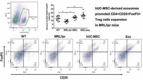
Previous studies have implicated that the transplantation of human umbilical cord mesenchymal stem cells (hUC-MSCs) effectively alleviates systemic lupus erythematosus (SLE) primarily due to immunomodulatory effects. However, little is known about the role of hUC-MSC-derived exosomes in SLE. This study is carried out to investigate the modifying effects of hUC-MSC-exosomes on the differentiation and function of immune cells in SLE. hUC-MSC-derived exosomes were extracted from the cultural supernatant of hUC-MSCs by ultrahigh speed centrifugation. Quantitative real-time polymerase chain reaction, western blot, enzyme-linked immunosorbent assay, and flow cytometry were performed to estimate the effect of hUC-MSC-derived exosomes on macrophage and regulatory T cell (Treg) polarization. In vivo, hUC-MSC-exosomes were injected intravenously into 28-week-old MRL/lpr mice. We had found that exosomes derived from hUC-MSC restrained the proliferation and inflammation of macrophages in vitro. Besides, MSC-exosomes inhibited CD68+M1 and HLA-DR+M1 but promoted CD206+M2 and CD163+M2 in vitro. Moreover, MRL/lpr mice administrated by intravenous injection of MSC-exosomes had less infiltration of CD14+CD11c+M1 cells but more CD14+CD163+M2 cells as well as Tregs in spleens compared with those in MRL/lpr mice treated by PBS. Additionally, MSC-exosomes could alleviate nephritis, liver and lung injuries of MRL/lpr mice. The survival of lupus mice could be improved after MSC-exosome treatment. This study has suggested that MSC-derived exosomes exert anti-inflammatory and immunomodulatory effects in SLE. MSC-exosomes ameliorate nephritis and other key organ injuries by inducing M2 macrophages and Tregs polarization. As natural nanocarriers, MSC-exosomes may serve as a promising cell-free therapeutic strategy for SLE.
Abbreviations: SLE: Systemic lupus erythematosus; hUC-MSCs: Human umbilical cord mesenchymal stem cells; MSCs: Mesenchymal stem cells; qRT-PCR: Quantitative real-time polymerase chain reaction; ELISA: Enzyme-linked immunosorbent assay; Tregs: Regulatory cells; TNF-α: Tumor necrosis factor alfa; IL: Interleukin; COVID-19: Coronavirus disease 2019; pTHP-1: PMA-induced THP-1 macrophages; TEM: Transmission electron microscopy; LPS: Lipopolysaccharide; EVs: Extracellular vesicles; TRAF1: Tumor necrosis factor receptor-associated factor 1; IRAK1: Interferon-α-interleukin-1 receptor-associated kinase 1; NF-κB: Nuclear factor-κB; BLyS: B lymphocyte stimulator; APRIL: A proliferation-inducing ligand
Graphical Abstract
Acknowledgments
We thank professor Jiyu Ju very much for his kind donation of MRL/lpr mice.
Authors’ contributions
WS, DX, SY and XL: conception and design, data analysis and interpretation, and manuscript writing. ZG, CY, JY, WS, SY, CL: collection and assembly of data, data analysis and interpretation, manuscript writing and revising. DX, XL, SY, WS, Lu Z and MC: manuscript revising. LZ, CY, JD and JY: collection and/or assembly of data and data analysis, and manuscript writing. ZG, SY, WS, and LZ: collection and/or assembly of data and data analysis, study material and interpretation. All the authors have read and approved the final manuscript.
Disclosure statement
No potential conflict of interest was reported by the author(s).