Abstract
A 14-year old girl was diagnosed as suffering from an immature teratoma. As removal of the teratoma was technically unfeasible, the patient was started on interferon α 2β, 3 MU/day, 5 days per week. On this therapy, the growth of the teratoma was stopped and the patient has been well for the past 50 months. She continues to be treated with interferon α 2β, with almost no side effects and no restrictions of her everyday activities, with a good quality of life. She is able to attend school regularly, travels abroad frequently, and participates fully in everyday activities normal for her age and social conditions. Her Karnofsky performance status is 100. This case demonstrates the efficacy of interferon α 2β therapy in certain instances of growing teratoma syndrome, even when the tumorous mass is initially quite large.
Keywords:
Germ cell tumors (GCT) of the ovary in childhood comprise only about 1% of all pediatric cancers Citation[1]. The general prognosis of such tumors is usually very good, with an over 80% 5-year disease-free survival Citation[2, 3]. Even when a patient presents with metastatic disease, the chances of cure are good with modern therapy which combines chemotherapy, radiation therapy, and surgery Citation[2]. Sometimes, successful treatment of GCT is accompanied by the appearance of secondary masses consisting histologically of benign cells. This phenomenon has been designated as growing teratoma syndrome (GTS) Citation[4]. GTS may appear in various parts of the body (retro- and intra-peritoneum, mediastinum, brain). Definitive treatment of GTS is complete surgical resection of the tumorous mass when feasible Citation[5–7]. Problems arise when definitive surgery is technically impossible and the continuous growth of the tumor begins to potentially endanger the health and life of the affected person. We describe a patient with unresectable GTS of the abdominal cavity, treated with interferon α 2β with resultant stabilization of the growth of the tumor for a prolonged period of time.
CASE REPORT
A 14-year-old girl of previously excellent health was first admitted to another hospital in December 1998 with complaints of severe abdominal pain. In the Emergency Room, the suspicion of a ruptured ovarian cyst was raised. The patient underwent left adnexectomy, and her postoperative period was uneventful. Histologic examination of the removed ovary revealed no malignant tissue and the pathologist concluded that it was a dermoid cyst. Two months later, the patient was reevaluated because of an increase in abdominal girth. Computer tomography (CT) scan of the abdomen revealed a huge non-homogenous mass, 10×8×12 cm, with displacement of the uterus and significant ascites as well as multiple tumorous nodules scattered throughout the entire abdominal cavity. The level of alpha-fetoprotein (α -FP) was significantly increased (above 1000 ng/mL). Review of histologic slides obtained at the time of her previous surgery demonstrated immature teratoma, grade 0–I, with positive staining for α -FP.
The patient was treated with two courses of BEP (bleomycin 30 mg/m2, etoposide 500 mg/m2, and cisplatin 100 mg/m2) chemotherapy without any apparent effect on tumor growth, and it was decided to proceed to a second-look operation. In March 1999, she underwent resection of a primary tumor, hysterectomy, right ovariectomy, and omentectomy. A week later, she was given cisplatin 50 mg intraperitoneally and referred to our service. As she was initially treated in a hospital abroad, we do not have sufficient information about the reasons behind these therapeutic decisions.
On admission to our hospital, the patient was in good clinical condition, without signs of distress. On physical examination, there was significant enlargement of the abdomen, with protrusion of its lower part due to pressure of the tumor on the abdominal wall. The following work-up was performed:
CT scan of the abdomen: large abdominal mass originating most probably from the pelvis and multiple tumorous nodules scattered within the entire abdominal cavity, along with significant ascites
CT scan of the chest: normal
Isotopic investigation with Tc99m: normal
α-FP and β-hCG blood levels: normal
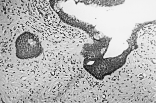
Multiple biopsies from several tumor sites and peritoneal nodules were taken, with histology showing only a mature teratoma (). It was concluded that the patient was suffering from so-called growing teratoma syndrome. Because of the widespread nature of the tumor and its proximity to various internal organs (bladder, intestines), definitive surgery was not feasible. Chemotherapy did not seem justifiable, and it was decided to treat the patient with interferon α 2β. In July 1999, she was started on Roferon (Roche Pharmaceuticals Ltd, Switzerland) 3 MU/day, subcutaneously, 5 times a week. A mere 2 weeks after starting this therapy, progression of the increase in abdominal girth stopped.
1 Mature epithelial nests and cysts, containing squamous, respiratory, and simple epithelia, embedded within mature fibrous mesenchymal tissue (H&E, ×200).

On repeated CT scans of the abdomen, other than an increase in the number of intra-abdominal calcifications, no further growth of the abdominal tumor was detected for the subsequent 11 months. During this time, our patient was well, continuing regular attendance at school and participating in all the usual activities for a child of her age.
In October 2000, it was decided to perform partial resection of her teratoma because of the significant psychological distress experienced by the girl due to her protuberant abdomen. Surgery was uneventful and, after surgery, another drug with prominent antiangiogenetic properties, tamoxifen, was added at a rate of 60 mg/m2/day. The decision to add tamoxifen was based on reports investigating this drug as an antiangiogenetic agent in combination with interferon-beta in conditions of various sold tumors Citation[8]. We were aware of potential angiogenesis stimulation which might have occurred immediately after abdominal surgery, caused by the release of various substances (vascular endothelial growth factor, platelet-derived growth factor) by the tumor as a result of its partial resection. Levels of α -FP and β -hCG were within normal range during the interferon therapy (< 2 ng/mL and < 5 ng/mL, respectively).
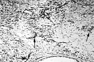
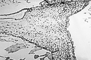
After partial resection was performed, pathological tissues obtained during the first exploratory laparotomy in May 1999 and during the partial resection of the teratoma in October 2000 were stained for von Willebrand's factor to ascertain the possible antiangiogenetic effect of interferon α 2β. A significant decrease in intensity of staining was demonstrated over the course of time (). The vessel density was determined by counting the number of vessels in 10 high-power fields in the areas with the highest number of vessels. Blood vessel count was evaluated following endothelial staining using an antibody to von Willebrand's factor (polyclonal; dilution 1:50; Zymed Laboratories Inc., South San Francisco CA). Although von Willebrand's factor staining is not specific for endothelial cells, in this particular setting, combined with histologic features, the stain highlights the blood vessels (which are recognized by their typical histology).
2 Many stromal capillaries highlighted by staining for von Willebrand's factor (bottom, gland with mucinous epithelium).

3 Most of the stromal area is deficient in capillaries (some are present in the upper area of the stroma). Stain for von Willebrand's factor.

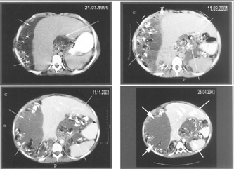
In October 2001, the girl developed the clinical picture of a partial bowel obstruction. Conservative methods of treatment did not provide resolution of symptoms (abdominal pain, vomiting) and she underwent exploratory laparotomy with dissection of bowel adhesions. Since then, she has continued treatment with interferon α 2β and tamoxifen, living a completely normal life, with regular attendance at school and without any side effects of her treatment. CT of her abdomen performed during the course of her illness (from July 1999 to April 2003) has demonstrated a stable size of teratoma ().
4 CT scans with contrast enhancement obtained before initiation of interferon therapy and during the course of interferon therapy, showing from moderate to significant amounts of ascites flanking the liver and spleen. Note some tiny calcifications and a few foci of soft tissue attenuation within the peritoneal fluid.

DISCUSSION
In 1982, Logothetis et al. Citation[4] first described the clinical phenomenon of the local growth of a histologically benign tumor in six men who had enlarged neoplastic masses after therapy for germ cell cancer. The clinical definition of what has been called ‘growing teratoma syndrome’ includes several components, including a history of non-seminomatous germ cell tumor (NSGCT) with teratomatous tissue in the primary specimen, along with elevated tumor markers, such as α -FP, β -hCG, and LDH, normalization of these markers after successful therapy, and enlargement of the metastatic tumor in the presence of normal tumor markers Citation[4]. Most cases of this syndrome have been reported in male patients suffering from NSGCT Citation[9, 10]. The growing teratoma can be located in various sites; this syndrome has been described in patients with GTS in the brain Citation[11], mediastinum Citation[12], and, most commonly, in the abdominal cavity Citation[5, 13–15]. In the vast majority of cases, GTS consists of only benign, mature tissue Citation[4, 5, 7, 15] and all signs and symptoms of the tumor are due exclusively to local pressure on surrounding structures Citation[14]. In very rare instances, the growing teratoma may contain malignant cells along with benign tissue. In most cases, these cells are comprised of endodermal sinus tumor (yolk sac tumor) with an ability to be stained positively for α -FP Citation[16, 17]. Elevation of a previously normal levels of α -FP in a patient with GTS proclaims malignant metamorphosis of the tumor and confers a poor prognosis Citation[18, 19]. Therefore, serial determination of α -FP levels is used for follow-up of these patients.
The definitive treatment of GTS is radical surgery where feasible Citation[5–7]. In their series of 11 patients, Maroto et al. Citation[5] reported that all patients who underwent radical removal of their teratoma survived without recurrence. In contrast, those whose growing teratomas were not completely resected had recurrences. Repeated and sometimes extensive surgery may lead to mutilation of such patients, causing significant morbidity. Our patient underwent two major operations, leaving her without her uterus and ovaries, requiring replacement estrogen therapy for the rest of her life. Given the benign nature of the growing teratoma and its frequent recurrence, we believe that too heroic efforts to remove all tumorous tissues should be avoided, and surgery should be performed only when there is direct danger to adjacent structures (e.g., obstruction of the gastrointestinal or urinary tracts) or when removing the tumor is feasible without endangering the life of the patient. In our patient, the repeated surgeries could have contributed to the stabilization of the tumor's size, but these were not the main reason for the stabilization, as repeated abdominal CT investigations revealed no further progression of the patient's teratoma throughout the 4 years of follow-up.
The mechanism and stimuli that cause uncontrollable teratoma growth are still unclear. Hormonal and cytokine stimulation have been proposed Citation[16]. The fact that most GTS patients are young men and women, frequently adolescents, when the sex hormone status is undergoing significant change, renders the theory of hormonal imbalance as a cause of GTS especially attractive. However, we found no reports in the literature showing any particular hormonal therapy as effective in the treatment of such patients.
Based on a few reports in the literature Citation[9, 14, 20, 21], we decided to treat our patient with interferon α 2β and the result was satisfactory. Other authors have also reported positive results with this kind of treatment, and some patients treated in this manner benefited from stabilization and even experienced a decrease in the size of the teratoma Citation[14, 20, 21]. There is one report of the complete disappearance of the growing teratoma in a patient who suffered from GTS in the abdominal cavity Citation[9]. Our patient is noteworthy in that stabilization of growth of her teratoma was achieved by interferon α 2β, despite the huge size of the tumor and multiple secondary implants in the peritoneum. Various chemotherapeutic agents are able to exert antitumor activity through the inhibition of angiogenesis. Our patient received two courses of BEP chemotherapy. Nevertheless, her teratoma had continued to grow rapidly, and it was concluded that, in this particular case, the BEP regimen failed both as a chemotherapeutic and an antiangiogenetic modality.
The exact mechanism of action of interferon in patients with GTS is unclear. It is believed that the interferon exerts its effect both through its direct antiproliferative and antiangiogenetic abilities and by its immune modulatory effect Citation[22]. Rustin et al. were the first to demonstrate the efficacy of interferon α in patients with mature teratoma Citation[20]. Two of their patients with differentiated teratomas, which can be now classified as GTS, responded to therapy with interferon.
In conclusion, patients with GTS not amenable to radical resection represent a difficult therapeutic problem. In such a situation, treatment with interferon may be justified in an attempt to stabilize, or even decrease, the size of the tumor. In situations when interferon therapy is ineffective, or where there are signs of impending obstruction of adjacent organs by the tumor, partial resection of the growing teratoma is indicated.
The authors wish to thank Mrs. M. Perlmutter for her assistance in the preparation of this paper.
REFERENCES
- Ries L, Smith M, Gurney J C, et al, Cancer incidence and survival among children and adolescent: United States SEER program 1975–1995. Bethesda, MD, National Cancer Institute, SEER program. NIH, (Pub. No. 99–1999;1–15).
- Gadducci A, Cosio S, Muraca S, Genazzani A R. The management of malignant nondysgerminomatous ovarian germ cell tumors. Anticancer Res.. 2003; 23: 1827–1836. [PUBMED], [INFOTRIEVE]
- Tewari K, Cappuccini F, Disaia P J, Berman M L, Manetta A, Kohler M F. Malignant germ cell tumors of the ovary. Obstet Gynecol.. 2000; 95: 128–133. [CROSSREF], [PUBMED], [INFOTRIEVE]
- Logothetis C J, Samuels M L, Trindale A, Johnson D E. The growing teratoma syndrome. Cancer. 1982; 50: 1629–1635. [PUBMED], [INFOTRIEVE]
- Maroto P, Tabernero J M, Villavicencio H, et al, Growing teratoma syndrome: experience of a single institution. Eur Urol.. 1997; 32: 305–309. [PUBMED], [INFOTRIEVE]
- Jeffery G M, Theaker J M, Lee A H, et al, The growing teratoma syndrome. Br J Urol.. 1991; 67: 195–202. [PUBMED], [INFOTRIEVE]
- Ravi R. Growing teratoma syndrome. Urol Int.. 1995; 55: 226–228. [PUBMED], [INFOTRIEVE]
- Lindner D J, Borden E C. Effects of tamoxifen and interferon-beta or the combination on tumor-induced angiogenesis. Int J Cancer. 1997; 71: 456–461. [CROSSREF], [PUBMED], [INFOTRIEVE]
- Ornadel D, Wilson A, Trask C, Ledermann J. Remission of recurrent mature teratoma with interferon therapy. J Roy Soc Med.. 1995; 88: 533–534
- Coscojuela P, Llauger J, Perez P, et al, The growing teratoma syndrome: radiologic findings in four cases. Eur J Radiol.. 1991; 12: 138–140. [CROSSREF], [PUBMED], [INFOTRIEVE]
- O'Callaghan A M, Katapodis O, Ellison D W, et al, The growing teratoma syndrome in a nongerminomatous germ cell tumor of the pineal gland: a case report and review. Cancer. 1997; 80: 942–947. [CROSSREF], [PUBMED], [INFOTRIEVE]
- Afifi H Y, Bosl G J, Burt M E. Mediastinal growing teratoma syndrome. Ann Thorac Surg.. 1997; 64: 359–362. [CROSSREF], [PUBMED], [INFOTRIEVE]
- Geisler J P, Goulex R, Foster R S, Sutton G P. Growing teratoma syndrome after chemotherapy for germ cell tumors of the ovary. Obstet Gynecol.. 1994; 84: 719–721. [PUBMED], [INFOTRIEVE]
- Kattan J, Droz J P, Culine S, et al, The growing teratoma syndrome: a woman with non-seminomatous germ cell tumor of the ovary. Gynecol Oncol.. 1993; 49: 395–399. [CROSSREF], [PUBMED], [INFOTRIEVE]
- Tongaonkar H B, Deshmane V H, Dalal A V, et al, Growing teratoma syndrome. J Surg Oncol.. 1994; 55: 56–60. [PUBMED], [INFOTRIEVE]
- Gobel U, Calaminus G, Engert J, et al, Teratomas in infancy and childhood. Med Pediatr Oncol.. 1998; 31: 8–15. [CROSSREF], [PUBMED], [INFOTRIEVE]
- Heifetz S A, Cushing B, Giller R, et al, Immature teratomas in children: pathologic considerations. Am J Surg Pathol.. 1998; 22: 1115–1124. [CROSSREF], [PUBMED], [INFOTRIEVE]
- Ahlgren A, Simrell C, Triche T, et al, Sarcoma arising in a residual teratoma after cytoreductive chemotherapy. Cancer.. 1984; 54: 2015–2018. [PUBMED], [INFOTRIEVE]
- Ulbright T, Loehrer P, Roth L, et al, The development of non-germ cell tumors: a clinicopathologic study of 11 cases. Cancer. 1984; 54: 1824–1833. [PUBMED], [INFOTRIEVE]
- Rustin G JS, Kaye S B, Williams C J, et al, Response of differentiated but not anaplastic teratoma to interferon. Br J Cancer. 1984; 50: 611–616. [PUBMED], [INFOTRIEVE]
- Van der Gaat A, Kok T, Splinter T. Growing teratoma syndrome successfully treated with lymphoblastoid interferon. Eur Urol.. 1991; 19: 275–278
- Taylor-Papadimitriou J, Ebsworth N, Rozengurt E. Possible mechanisms of interferon-induced growth inhibition. Symp Fundam Cancer Res.. 1984; 37: 283–298. [PUBMED], [INFOTRIEVE]