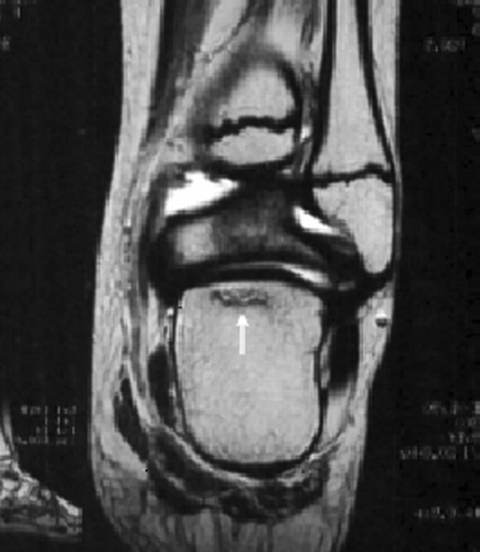
We have read the interesting report by Dr. Goldbloom et al. regarding osteoporosis at presentation of childhood ALL. We would like to present the case of an adolescent girl with ALL who developed osteoporosis-related complications after chemotherapy and was successfully treated with alendronate.
A 10-year-old girl was admitted to our department with a history of pain and swelling in the left knee for 1 month. Diagnosis of T-lineage ALL was confirmed by bone marrow aspirate. The girl was treated with high-risk ALL BFM'95 protocol. Bone marrow examination at 33 days confirmed a complete remission. Evaluation of bone metabolism with bone mineral density (BMD; g/cm2) of lumbar spine (LS) and femoral neck (FN) at diagnosis was normal (z scoreLS = −0.25, z scoreFN = −0.1) but after induction chemotherapy revealed severe osteopenia (z scoreLS = −1.95, z scoreFN = −2.33) and she was started treatment with calcium carbonate and alfacalcidol. Two months later, while on consolidation treatment, she complained of severe pain in the right ankle joint, only during walking. Radiographs of the right foot revealed diffuse osteoporosis and diagnosis of astragalus osteonecrosis was posed after magnetic resonance images (). She was temporarily started on a conservative medical treatment consisting of physical inactivity and use of a pair of crutches for pain reduction. She was evaluated after 1 month, and MRI images indicated amelioration. On a periodic control (3 months later) with lumbar spine and femoral neck BMD measurement, the girl continued to be osteopenic (z scoreLS = −2.01, z scoreFN = −2.24). Two months later while on consolidation treatment she presented back pain without history of injury and X-ray and MRI scans of lumbar spine showed a collapse of the vertebral body (T-9). She was treated with alendronate 70 mg (once a week), calcium, physical therapy, and orthopedic corset. Recent radiological examinations showed a mild recovery of the vertebral body collapse at 3 and 6 months follow-up. Moreover, 6 months after the initiation of treatment with biphosphonates, the patient's z score was ameliorated (z scoreLS = −1.33, z scoreFN = −1.51).
Many studies in literature have demonstrated that children affected by ALL are osteopenic/osteoporotic at all phases: at diagnosis, during treatment, and mainly after completion of treatment [Citation[1], Citation[2]]. The pathogenesis of osteopenia–osteoporosis is multifactorial and the main risk factors include leukemic invasion of the bone at diagnosis, corticosteroid and methotrexate treatment, cranial irradiation, hormone deficiencies, malnutrition, and prolonged physical inactivity. Furthermore, many studies in the literature have shown that bone loss after long corticosteroid administration is most rapid in trabecular bone. In our patient BMD at both sites (LS and FN) was indicative of severe osteopenia. Disturbed bone metabolism due to corticosteroids (mainly dexamethasone) in our patient might have been the main factor responsible for the development of osteonecrosis. Moreover, osteopenia was aggravated by physical inactivity after osteonecrosis and was another contributing factor for developing of the vertebral fracture.
The incidence of musculoskeletal manifestations and bone lesions after chemotherapy of childhood ALL is relatively high, but only a few cases of severe osteopenia–osteoporosis, osteonecrosis, and vertebral collapse secondary to treatment have been reported in the same patient [Citation[3], Citation[4]]. Although bisphosphonates have been used in adults for more than 30 years, their use in children has been limited. A recent study has reported a clinical improvement and a gradual increase in BMD in 10 children with ALL and NHL Citation[5]. We conclude that a follow-up of BMD could facilitate the identification of those children who may require specific therapeutic treatment such as biphosphonate to prevent any further decrease in their skeletal mass. Moreover, the use of bisphosphonate therapy could represent an additional strategy for optimization of bone mass and treatment of skeletal complications in children with hematological malignancies.
REFERENCES
- Goldbloom E, Cummings E, Yhap M. Osteoporosis at presentation of childhood ALL. Pediatr Hematol Oncol 2005; 22: 543–550, [INFOTRIEVE], [CSA]
- Haddy T, Mosher R, Reaman G. Osteoporosis in survivors of acute lymphoblastic leukemia. The Oncologist 2001; 6: 278–285, [INFOTRIEVE], [CSA], [CROSSREF]
- D'Angelo P, Conter V, Chiara G D, et al. Severe osteoporosis and multiple vertebral collapse in a child during treatment for B-ALL. Acta Haematol. 1993; 89: 38–42, [INFOTRIEVE], [CSA]
- Kakihara T, Watarabe A, Imai C, Hotta H, Tanaka A, Uchiyama M. Chemotherapy-induced multiple vertebral compression fractures in a boy with acute lymphoblastic leukemia. Pediatr Int 2002; 44: 683–685, [INFOTRIEVE], [CSA], [CROSSREF]
- Wiernikowski J T, Barr R D, Webber C, Guo C Y, Wright M, Atkinson S A. Alendronate for steroid-induced osteopenia in children with acute lymphoblastic leukaemia or non-Hodgkin's lymphoma: results of a pilot study. J Oncol Pharm Pract 2005; 11: 51–56, [INFOTRIEVE], [CSA], [CROSSREF]
