

Abstract
Bone defect restoration remains challenging in orthopedic medical practices. In this study an attempt is carried out to probe the use of new biomimetic SPEEK (sulfonated polyether ether ketone) based nanofibrous scaffold to deliver amine functionalized hydroxyapatite nanoparticles loaded resveratrol for its potent functionality in osteogenic differentiation. SPEEK polymer with reactive functional group SO3H was synthesized through process of sulphonation reaction. Amine functionalized nanoparticles with protonated amino groups revamp the molecular interaction by the formation of hydrogen bonds that in turn intensify the bioactivity of the nanofibrous scaffold. Osteoconductive functionalized nanohydroxyapatite enhances the cell proliferation and osteogenicity with improved cell attachment and spreading. The results of FT-IR, XRD, Carbon-Silica NMR and EDX analysis confirmed the amine functionalization of the hydroxyapatite nanoparticles. Surface morphological analysis of the fabricated nanofibers through SEM and AFM analysis shows vastly interconnected porous structure that mimics the bone extracellular matrix, which enhances the cell compatibility. Cell adhesion and live dead assay of the nanoscaffolds express less cytotoxicity. Mineralization and alkaline phosphatase assay establish the osteogenic differentiation of the nanofibrous scaffold. The in vitro biocompatibility studies reveal that the fabricated scaffold was osteo-compatible with MG63 cell lines. Hemocompatibility study further proved that the designed biomimetic nanofibrous scaffold was highly suitable for bone tissue engineering. The results of in vivo analysis in zebrafish model for the fabricated nanofibers demonstrated significant increase in the caudal fin regeneration indicating mineralization of osteoblast. Thus, the commending results obtained instigate the potentiality of the composite nanofibrous scaffold as an effective biomimetic substrate for bone tissue regeneration.
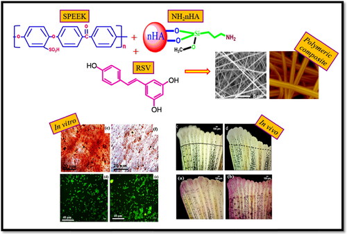
GRAPHICAL ABSTRACT
Acknowledgments
The authors would like to acknowledge the Indian Council of Medical Research (ICMR), New Delhi, India, for the financial support (Vide letter No. No: grant No. 45/47/2019-NAN-BMS, letter dated 30.09.2019). Authors would like to thank Dr. E. Shoba, CSIR-CLRI, Adyar, Chennai and India for providing support to carry out in vitro studies.
Disclosure statement
Authors declare that there is no conflict of interest.