Abstract
Crystalline characteristics of natural and acid modified starches (cereals, millets, pulses, root, and tuber) were examined by X-ray diffraction studies. The (hkl) planes were identified using A-, B- and C- type starch cell parameters. The size of crystallite along the direction [hkl] was calculated for all the reflections in the starches. Smallest crystallites (35 to 56 Å) are observed for the reflections at (2θ) 11.2 to 11.5° corresponding to (111) reflection plane, waxy rice showing the lowest value of 35 Å. Largest crystallite size of the order 200 Å is observed at 2θ 20.0 to 20.4° corresponding to (032) reflection plane with finger millet giving the highest value in the order of 289 Å. Acid modification leads to minor changes in ‘d' spacing but crystallite size changes quite considerably. It increases in some, decreases in others, and remains unchanged in few cases. There was no uniform pattern observed either with respect to acid used, source of starch or the peak at which the changes occur. In finger millet starch, crystallite size becomes homogeneous upon acid treatment.
INTRODUCTION
Starch has been regarded as a semi-crystalline material since the classic work of Katz in the 1930s using X-ray diffraction techniques. He has categorised 3 types of crystalline structures for the intact starch granules which gave three characteristic diffraction patterns designated as the A-, B- and C- types.[Citation1] The crystalline type depends on the botanical source of the starch: A-pattern, which is typically given by cereal starches; B-pattern by potato, amylo maize, canna, retrograded starch; and C-pattern by pulses, tapioca, arrowroot and sago.[Citation1–3]
The X-ray diffraction patterns could be used therefore to differentiate between various native starches. X-ray patterns can also detect changes in crystallinity brought about by physical or chemical treatments. The interplanar ‘d' spacing and the size of crystallites at different peak zones have been studied to understand the nature of the starch granule organization. Transmission electron microscopy studies have provided useful data on the nature of micro-granular structure and electron dense regions in starches. Kreger informed that a spiral configuration with 3 glucose residues per turn, which not only explains the fibre period, but also the hexagonal ratio of the axes in the basal plane of the provisional unit cell.[Citation4] He also conveyed that spiral chains could not be packed in the provisional unit cell. On account of hexagonal ratio of the axes in the basal plane a hexagonal unit cell is suggested with a = 18 A° and b = 10.6 A°, through which 18 chains runs i.e the cell contains 54 glucose residues and water molecules. The x-ray densities of such a packing is consistent with observed densities of starch grains (granules).[Citation4] Micelle dimensions for potato starch in a and b directions of Rundle's unit cell were estimated to be D(100) = 147 A and D (020) = 144A, respectively.[Citation5] On acid modification of waxy, high amylose maize, tuber, and tobacco leaf, electron microscope of corroded granules showed that acid attack was more rapid in certain parts of each concentric granule shell. It was well defined in acid degraded maize starches compared to tuber degraded starch. This was also confirmed that iodine colour with tuber starch faded fast compared to iodine colour of acid treated maize starches. All this, indicated that cereal starches differ from tuber and tobacco leaf starches.[Citation6] The micro-granular structure has been observed to be accentuated by enzymic or acidic erosion.[Citation7,Citation8] The technique has been employed by a number of workers and starch soaked in acid of dilute or moderate concentration over a period of time (Lintnerization) has been examined for granular and molecular properties.
Sarko and Wu[Citation9] studied the crystal structure of A-, B-, and C-polymorphs of amylose by the combination of X-ray diffraction and computer based structure refinement, and concluded that A and B structures differ in the crystalline packing of the helices and the water content and C-structure is a mixture of A and B unit cells and therefore intermediate between A and B forms in packing density.[Citation9] Nara et al. have reported that potato starch diffraction pattern becomes sharp with increase of sorption moisture and areas of crystallinity are proportional to the amount of moisture.[Citation10] Fine rice flour (Basmati 370) and coarse rice flour (Jaya cultivar) showed A type diffraction pattern but coarse showed more crystalline than fine flour. Mixing these flour with some of legume flours did not change much in ‘d' spacing and hence they were characterized as A type. On extrusion of blends, A type changed to ‘V' type indicating lipid amylose complex formation during extrusion.[Citation11] Granule size and shape, apparent amylose, reducing value, Differential scanning calorimetry and X-ray diffraction of two curcuma starches were studied. X-ray diffraction was of B type and many differences were not seen. The peaks for these two starches were observed at 2 of 15 to 18.6 and 23 to 26.[Citation12] Even native starches from various sources were acid hydrolysed and compared with the enzyme hydrolysis, degradation pattern were studied and concluded that enzyme hydrolysis was quite fast.[Citation13] The present paper reports data on the crystalline nature of various starches and the effect of acid modification on crystal parameters.
MATERIAL AND METHODS
Maize and tapioca starches were procured from commercial sources. Potato starch was obtained from Professor W. Kempf of Federal Research Center for Cereal and Potato Processing, Detmold, Germany. Chickpea and green gram pulses were procured from the local market. All chemicals used were of analytical grade. Starches from all other source materials (procured from local market) were isolated in the laboratory as reported elsewhere.[Citation14–16] In the procedure, the starches were acid modified (33% starch slurry) using 0.5 N HCl, HNO3, H2SO4 and H3PO4 for 1.5 h at 50°C, with occasional stirring. At the end, they were neutralized with NaOH and washed to free from respective anions. The starches were dried and ground using Fritsch pulverizer with 2.0 mm screen. Samples had moisture content of about 12.5% (w.b) and protein content between 0.1 and 0.3% (w.b) estimated by Kjeldhal method (N × 6.25).
X-ray Diffraction Pattern
X-ray diffraction spectra were obtained using a Phillips X-ray Diffractometer system PW 1710 equipped with a mono chromator that selects Kα radiation from a copper target generated under 35 kV (target voltage), 15 mA (tube current), Phillips, Netherlands. The spectra were recorded at a scan speed of 2.4°/min. having a width of 1° of 2θ for 1 cm. The scanning angle range of 2θ was 5° to 30° for native as well as modified starches.
Analysis of X-ray Diffractogram
-
For measuring the crystallinity of starch from the diffractograms, a base line was drawn from starting of the curve to the end of the curve as well as at the base of peaks. The area of upper region above peak base line, representing X-ray scattering of the crystalline portion, as a proportion to the whole area under diffractogram gave the index of crystallinity.
(1)
-
Diffraction spacing ‘d,' i.e., interplanar distance for each peak was calculated using the Bragg's equation
where λ = 1.5418 Å, n = 1 (first order reflection), θ = the Bragg angle for corresponding peak.
X-ray reflections were identified in terms of Miller indices (hkl) by trial and error[Citation17] computer program. Crystal size D was calculated using Scherrer equation.[Citation18]
RESULTS AND DISCUSSION
Peak Assignment
The X-ray diffractograms of six starches viz. of maize, finger millet, wheat (cereals), green gram (pulse), tapioca (root), and tuber (potato), both for native and modified starches using different acids are presented in . compares the assigned 2θ values of different X-ray types (A, B and C) as per literature[Citation2] with those observed for the starches except for a few minor differences, the peaks assigned in literature appear in our diffractogram. The very weak (W) peak around 10° reported in the literature is absent in all our starches. However, a close examination of the diffractograms reveals that the peak is rather ambiguous and perhaps could become pronounced had the starches been hydrated to a higher moisture content (about 25%, by equilibrating in humid air).[Citation2]
Figure 1 X-ray diffractograms of selected starches before and after modification With acids. 1 = Native, 2 to 5, modified with HCl, HNO3, H2SO4 and H3PO4, respectively (0.5 N acid, 50°C, 1.5h).
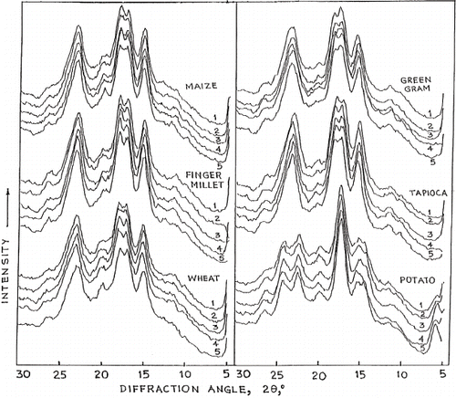
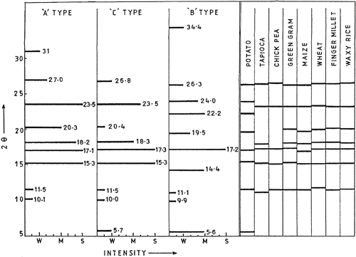
Figure 2 Schematic Intensity plots at specific 2θ values of A, B and C type starches as per CitationZobel, 1964. Intensity scale: Weak (W), Medium (M), and Strong (S). The observed 2θ values for starches are indicated alongside for comparison.
The peak at 14.4° for potato starch is also present in the native but the intensity is not as strong as reported in the literature.[Citation2] Additionally, there is a peak at 15° not as strong in potato as in other starches. Acid modification pronounces the peak at 15° in some; maximum in H3PO4 modified potato starch. As a matter of fact, phosphoric acid increases the characteristic peaks assigned to ‘B' type starch, especially the one at 5.6°.
As for cereals, all the typical ‘A' type peak assignments are observed in the maize, wheat, rice, and finger millet starches studied, except that the peak at 27.0° appears at a lower angle viz. 26.2–26.5°. Tapioca and pulses, belong to ‘C' type X-ray diffraction pattern and possess partial characteristics from A and B types. With notable absence of peak at 5.73°, the peak at 20.3° or 20.4° appears very weak (W) and this agrees with literature reports. The peak at 18° is absent or ambiguous in chickpea while it is present in green gram. An examination of the diffractograms presented by Biliarderis also shows that this peak is absent in smooth pea, but present in adzukibean.[Citation19] The peak at 5.6° was however present in all the three legume starches studied by them. The minor differences among the peaks identified could be due to natural and varietal conditions and their very low moisture content (12.5%, w.b). Hydrated starches are known to give well-defined peaks, where added moisture permits the already longitudinal molecules in the amorphous regions to organize into crystalline units.[Citation20] The observed differences may fall within certain range of acceptability.
Miller Indices for Crystal Lattice
shows data on relevant crystal parameters of native and acid modified starches. Ambiguous peaks as discussed above, are excluded. The inter planar distance ‘d' for the observed peaks in the range of 5 to 30°, and size of crystallite at this spacing are presented. Miller indices for each reflection are identified using trial and error programme and for unit cell dimensions () assigned for A, B and C type starches reported by Sarko and Wu.[Citation9] These data are presented for various starches for the first time in the starch technology. The reflections may be grouped into 10 types in all. Reflections (001) and (113) are specific to potato starch at 5.6° and 22.2°, respectively, characteristic of ‘B' type. Reflections (140) for 15.2–15.5°, (113) for 17.0–17.4°, (132) for 23.2–23.9° and (142) for 26.2–26.5° appear for all starches. Reflection (111) for 11.2–11.80 is present in all except native and HCl-modified maize starches. Reflection (150) for 18.0–18.2° is absent for potato (as expected) and for chickpea. However, this reflection appears as a mild shoulder to that at 17° and is therefore ignored as ambiguous in chickpea. Reflection (032) expected at 20.0–20.4° is absent in potato, maize and chick pea starches.
Table 1 Diffraction spacing ‘d' and crystal size ‘D' of native and acid modified starches
Table 2 Unit cell dimension of A, B and C-type starchesFootnote∗
Crystallite Size (D)
The sizes of crystallites in the crystalline regions (peaks) of native and acid modified starches are calculated as per Scherrer equation (). For the native starches, the smallest crystallite size (average value: 42 Å) is for (111) reflection at 11.5°. Among different starches, the lowest value (35 Å) is for waxy rice, while the highest (56 Å) is for chickpea starch. Crystallite for (132) reflection at 23° has the next higher size (average: 90 Å), the lowest among them (75 Å) is for chick pea while the highest (116 Å) is for potato. The largest crystallite size is for (032) reflection, at 20°. The highest among them (289 Å) is for finger millet starch, followed by 269Å for waxy rice and 202Å for tapioca and green gram. The crystallites at 2θ equal to 15, 17, 18 and 26°give intermediate sizes, between 100 and 200 Å. Except for the early results of Hizukuri and Sterling, data on the crystallite size in starch are lacking.[Citation5] The former reported native potato starch crystallites having diameter 147 Å and 144 Å at 5.5 and 17.0°, respectively, while Sterling and Pangborn[Citation20] published values of 77 and 87 Å for native and Linternerized potato starch at 5.6°. These values are written 35 to 288 Å determined in the eight starches presented here. The crystallite size is different in different directions and varies from 35 to 288Å, indicating that shape of the crystallites is not definitely spherical.
Effect of Acid Modification
Acid modification brings about very small changes in the interplanar spacing ‘d' and considerable changes in crystallite size. There is no uniform pattern with respect to acid used, source of starch or peak position. The crystallite size increases in some starches, decreases in some others while stays put in the rest. There is an increase in the micellar diameter from 77 and 99 Å, for dry and wet potato starch, to 87 and 126 Å for dry and wet Lintnerized starch.[20] Crystallite for reflection (132) at 23°, shows the least change in size, with practically the same crystallite size irrespective of modification by any acid for tapioca, maize and chick pea starches. Wheat starch shows marginal decrease for HCl, H2SO4 and H3PO4 while green gram shows marginal increase for HCl and HNO3 treatment. Finger millet starch shows an increase in the crystal size as a result of modification by any acid. Additionally, all acids increase its D at 17° (131) and decrease those at 15° (140), 18° (150), and 20°(032). D is stabilized around 160 Å by appropriate adjustment of HCl and HNO3 modification. Thus, the D value of 134 Å, 201 Å, and 289 Å at 17, 18, and 20 in native finger millet starch become 161 Å for all the 3 peaks upon acid treatment. This trend exists in other starches also to a smaller extent. The variation in crystallite size caused by acid treatment could be due to disruption of amorphous region so that lateral organisation enhances, thus improving the overall crystallinity within the treated granule. This would sharpen the X-ray diffraction pattern.[Citation21] Thus, partial erosion of amorphous region and subsequent alignment of crystallites may increase its crystallinity. Against this, disruption but non-alignment may decrease the crystallite size.
Crystallinity of Starch
The overall relative crystallinity expressed in terms of area under peaks to that under complete diffractogram for each starch studied is given in for six native and modified starches. The lowest crystallinity (23%) is recorded for native potato starch. Tapioca has the highest crystallinity of 35%, while others are in between. Except for potato starch, which shows an increase of 4 to 8 percentage points in crystallinity, others are insensitive to acid modification. As the time of acid treatment (1.5 h) is short, this observation is understandable. Proneness of potato starch to rapid changes of crystallinity signifying its rather weak organization, is further seen in 13C CP/MAS NMR spectrums[Citation15], alkali fluidity number, molecular weight (Mn) upon storage of acid modified and not neutralized potato starch.[Citation14]
Table 3 Relative crystallinity (%) of starches before and after 0.5N acid modification
CONCLUSION
Data on the interplanar ‘d' spacing alone do not describe the crystalline properties of starch. However, characterisation of the reflections by the Miller indices (hkl) by using the unit cell dimensions reported by Sarko and Wu and the observed ‘d' spacings have enabled us to calculate crystallite size in the crystalline regions with the help of Scherrer equation.[Citation9,Citation18] Acid modification of starch increases the crystallite size in certain crystalline regions and decreases in others. There is no consistent pattern in the change. In finger millet starch, however, the crystallite size in the major crystalline region (at 17, 18 and 20°) changes to 160 Å irrespective of its earlier size (130 and 289 Å). It is evident from these results that crystallite shape is far from spherical shape. It is to be emphasised here that we have not included the para crystalline disorder, which may play dominant role in the crystallite shape and size.
Related Research Data
REFERENCES
- Katz , J.R. 1937 . Changes in X-ray Pattern When Starch Preparations are Dried, as a Way of Characterizing These Substances with X-ray . Recl. Tarav. Chim. Pays-Bas Belg , 56 : 766 – 772 .
- Zobel , H.F. 1964 . “ X-ray Analysis of Starch Granules ” . In Methods in Carbohydrate Chemistry, , IV , Edited by: Whistler , R.L. London : Academic Press Inc. .
- French , D. 1984 . Organisation of Starch Granules, Starch Chemistry and Technology , 2nd , Edited by: Whistler , R.L. , BeMiller , J. N. and Paschall , E. F. 183 – 247 . Academic Press Inc .
- Kreger , D.R. 1951 . Configuration and Packing of the Chain Molecules of Native Starch as Derived from X-ray Diffraction of Part of a Single Starch Grain . Biochim. Biophys. Acta , 6 : 406 – 425 .
- Hizukuri , S. and Nikuni , Z. 1957 . Micelle dimension of Potato Starch . Nature , 180 : 436 – 437 .
- Buttrose , M.S. 1963 . Electron Microscopy of Acid Degraded Starch Granules . Staerke/Starch , 15 : 85 – 93 .
- Nikuni , Z. 1978 . Studies on Starch Granules . Staerke/Starch , 30 : 105 – 111 .
- Chabott , J.F. , Allen , J.E. and Hood , L.F. 1978 . Freeze-ETCH Ultrastructure of Waxy Maize and Acid Hydrolysed Waxy Maize Starch Granules . J. Food Sci , 43 : 727
- Sarko , A. and Wu , C.H. 1978 . The Crystal Structure of A-, B- and C- Polymorphs of Amylose and Starch . Staerke/Starch , 30 : 73 – 78 .
- Nara , Sh. , Mori , A. and Komiya , T. 1978 . Study on Relative Crystallinity of Moist Potato Starch . Staerke/Starch , 4 : 111 – 114 .
- Chauvan , G.S. , Sharma , P and Bains , G.S. 2003 . Effect of Extrusion Cooking on X-ray Diffraction, Characteristics of Rice and Rice Legume Blends . Int. J. Food Prop , 6 ( 1 ) : 127
- Jyothi , A.N. , Moorthy , S.N. and Vimala , B. 2003 . Physicochemical and Functional Properties of Starch from Two Species of Curcuma . Int. J. Food Prop , 6 ( 1 ) : 1356
- Singh , V. and Ali , S.Z. 2006 . In vitro Hydrolysis of Starches by α – amylase in Comparison to That by Acid . American Journal of Food Technology , 1 ( 1 ) : 43 – 51 .
- Singh , V. and Ali , S.Z. 1987 . Comparative Acid Modification of Various Starches . Staerke/Starch , 39 : 402 – 405 .
- Singh , V. , Ali , S.Z. and Divakar , S. 1993 . 13C CP/MAS NMR Spectroscopy of Native and Acid Modified Starches . Staerke/Starch , 45 : 59 – 62 .
- Singh , V. and Ali , S.Z. 2005 . Properties of Starches Modified by Different Acids . Carbohydrate Polymers , (Submitted)
- Laugier, J. Powder Indexing Systems http://www.ccp14ac.in/tutorial/crys/index. html
- Glasstone , S. 1940 . “ Solid State ” . In Text Book of Physical Chemistry, , 1st 340 – 426 .
- Biliaderis , C.G. , Grant , D.R. and Vose , J.R. 1981 . Structural Characterisation of Legume Starches; II Studies on Acid Treated Starches . Cereal Chem , 58 : 502 – 507 .
- Sterling , C. and Pangborn , J. 1960 . Fine Structure of Potato Starch . Amer. J. Botony , 47 : 577 – 582 .
- Kainuma , K. and French , D. 1971 . Naegeli Amylodextrins and Its Relation to Starch Granule Structure. I. Preparation and Properties of Amylodextrin from Various Starch Types . Biopolymers , 10 : 1673 – 1680 .