Abstract
The microscopic structure of rough rice kernel has been investigated using Scanning Electron Microscope (SEM) after solar drying in order to identify the mechanisms of the initiation and propagation of stress induced cracks. The structure of the endosperm tissue of rice kernel has been found damaged slightly due to drying. Stress induced cracks were evident. The cracks were also found between the seed capsule and the endosperm tissue. The cracks appeared to have initiated at the center of endosperm tissue and then propagated. When stress induced cracks are produced, both sides of the endosperm tissue are damaged. Most of these cracks are propagated along the edge of the starch granule through to the cell walls. These observations are a part of a series of the studies which provide useful information for practical control or minimization of the crack formation in keeping the grade of the dried rice high.
INTRODUCTION
Stress induced cracks in rough rice usually refer to that cracks produced in the seed endosperm tissue, while the capsule tissue remains intact. These cracks, which weaken the rice mechanical strength, are often produced during harvest, drying, and storage. The price of the cracked rice is 1/3 less than the intact rice, so economic losses are heavy. After rice is cracked, it is easy to develop mould and pests. Because the endosperm tissue is broken, the nourishment that an embryo can receive is reduced. These factors reduce the vitality of the seeds.[Citation1]
Stress cracks of rice are traditionally observed using the light box method, which is dependent on many human factors such as mental state, vision state, and resolution capabilities of individuals. The observed results have a large variability. Using this method, only the surface cracks can be observed, but the location and propagation size of internal cracks of rice cannot be observed. In recent years, agricultural engineering experts have adopted many new physical and chemical methods, such as computer visualization method,[Citation2,Citation3] image processing method,[Citation4] neural network method,[Citation5] laser method,[Citation6] and so on, to determine the damages or the cracks of on and within rice. However, these methods are not popular due to their limited resolution compared with the rice kernel sizes. Again, the internal microstructure of rice cannot be observed by the previously-mentioned methods. At present, MRI and Scanning Electron Microscope (SEM) are the main technologies to observe the internal structure of rice. With MRI, the rice kernel can be scanned without damage to gain internal multilevel scanning image.[Citation6] Thus, the internal moisture distribution and location of stress induced cracks of the rice can be determined. Its disadvantage is that the microstructure of stress cracks cannot be established, due to its poor resolution.[Citation7] As such, SEM would be more appropriate for microstructure analysis.
In solar drying, rice kernel absorbs energy from solar radiation and internal moisture of rice is moved to the surface. Analysis and research on internal structure of rice after solar drying helps us understand the mechanisms of stress induced cracking and their propagation. In this article, we investigated the analysis the origin, propagating direction of the stress induced cracks, and the change of internal tissue structure of the rice after cracks were formed. This study will provide a good basis for future research for improving rice quality.
MATERIALS AND METHODS
The rough rice for experiment is the fresh rice harvested in the Guilin region of Guangxi Province, P. R. China. The variety is thus the “Gui 99.” After being threshed by hand, rough rice was put into the constant temperature incubator, in which the temperature was set at 0°C in order to keep the rough rice fresh. Experience shows that the storage for a short time at 0°C does not affect the quality of rough rice.[Citation8] Solar drying was carried out in the sunny and non-windy weather in autumn (September period in Beijing). The average temperature of air was about 20°C. The initial moisture content of rice was 23% (w. b.). Solar drying was stopped when the final moisture content reached 13% (w. b.). This lasted for 4 days. The intact and plump rice were picked randomly as the samples for post-drying analysis.
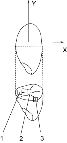
Traditional sampling method is that the samples need to be subdivided after being soaked, then drying them to the required moisture content.[Citation9] With this method, the samples would go through a wetting and then drying process. The extent of damage is usually enlarged. In order to avoid such a situation, in this study, the method of freezing using liquid nitrogen for sample preparation was employed. Once the sample rice particles were frozen, they can be gently knocked with a metal stick. The particles would start forming cracks under these very small forces. It has been found the majority of the rice granules start to crack forming crack lines circling around the surfaces parallel to the X axis as shown in .
The treated samples ware then adhered to the conducting resin and placed them into an ionic coating splashing machine (EIKO model, Ibaraki, Japan). Thin gold coating was applied for 2 hours. The thickness of splashed gold coating is usually about 20 nm. This complies with the standard SEM sample preparation procedure. The SEM machine was a HITACHI S-530 Scanning Electronic Microscope. Accelerating voltage of 25kV was used in the current experiments.
RESULTS AND DISCUSSION
Microstructure of the Endosperm Tissue
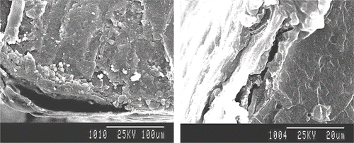
After drying, comparing the microstructure of internal tissue of rice with its initial microstructure, the difference was found to be inside the endosperm tissue. As shown in , the structure of endosperm tissue which was not damaged was very different from that has been some what damaged as shown in . Cell walls are apparent in , which are known to contain starch granules. shows some cells are in the process of being cracked through. The rupture of cell walls is taking place. Starch granules come from the cells. Some adjoining cell walls begin to separate. Stress induced cracks are produced.
Figure 2 Microscopic structure of the endosperm tissue after solar drying: (a) Structure of the endosperm tissue that was not damaged; and (b) Structure of endosperm tissue that was damaged slightly.
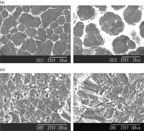
The structure of endosperm tissue of rice is damaged slightly after drying, because the rice kernels experienced temperature fluctuations in solar drying. Different structure strength can be found in different endosperm tissue cross sectional areas. As mentioned earlier, the rice was solar dried in the autumn period. It took 4 days for the moisture content to decrease from 23% (w. b.) to 13% (w. b.). The rice kernels went through 4 temperature cycles (day and night cycle). In this process, temperature gradient might be produced within the rice, and certain thermal stress was induced. The endosperm tissue could be subjected to torsion, compression and tension under the function of thermal stresses. In the end, cell walls could become weakened and sometimes become disintegrated. In the extreme case, starch granules would be “released” from within.
After drying, the microstructure of rice seed capsule was also observed using SEM. It is shown in . That seed capsule of rice is fiberous and should attach tightly against the inner tissue. It is not damaged as such, but the attachment between the seed capsule and the endosperm is more porous weaker. In certain areas, stress induced cracks were evident. This shows that the between seed capsule and endosperm can be easily separated, which may be mainly due to the expansion rate and the shrinking rate of the seed capsule was different from that of the endosperm. Repeated stressing cycles would induce damage between the seed capsule and the endosperm, in the long-time solar drying. In this way, the stress cracks were formed. It is also observed from that stress cracks between the seed capsule and endosperm is not directed to those of the endosperm. Embryo tissue was also observed using SEM. It was observed that from the micro-images, the cells of the embryo tissue were still arranged evenly and tightly, and there were no stress cracks or any sign of damage.
Microstructure of the Stress Cracks in the Endosperm
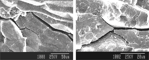
Using the light box method, we observed that the percentage of rice particles that have stress cracks after solar drying was only less than 3%. With SEM, there were stress cracks even inside the endosperm. The length and width of the cracks were small. Stress cracks in the endosperm are the cracks of limited lengths. They were distributed in endosperm without definite direction. Most are distributed in the seed core. The cracks were curving. When the stress cracks were produced, only the endosperm tissues of both edges of the cracks were damaged, but the tissues in between the cracks were kept intact. There were no smaller micro-cracks visible, as shown in .
Generation and Propagation of the Stress Cracks in the Endosperm
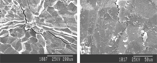
We observed stress cracks near the rice seed capsule using SEM. It is shown that the closer to the seed capsule, the stress cracks were narrower. Most stress cracks are distributed radiating from the center outwards, so the stress cracks were in fact produced in the area far from the seed capsule, and then propagated towards the seed capsule. They had, however, no capability to cut through the aleuronic layer, so the seed capsule remained intact as shown in .
After solar drying, the propagating routes of most stress cracks inside rice are those passing in between cells or may be said to cut through the cell wall regions, and then propagating along the edges of starch granules. But there are some other circumstances. for example, some stress cracks were found not only propagated along edges of starch granules, but also tore through some of the starch granules, dividing them into two pieces (as shown in ).
Figure 6 Microscopic structure of the stress cracks through the endosperm tissue after solar drying.
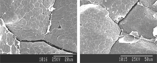
In solar drying, because the temperature of rice rises only slowly and the drying rate is low, the thermal stress, and the extent of shrinkage would be small. These factors would not heavily influence the network structures between starch granules and protein. The cells of the endosperm tissue of rice are bigger, with the shape of a polygon. Stress cracks are produced more easily when these cells are affected by mechanical stresses. The starch granules and protein “binding” structures would be relatively stronger. The cell walls were disintegrated and the stress cracks could propagate through them after solar drying. It proved that cell walls of the endosperm tissue of rice could stand little external stress.
CONCLUSION
After sun drying, endosperm tissues of rice were destroyed slightly and stress cracks were produced. Stress cracks were produced in a combination of areas between the seed capsule and endosperms of rice. But the stress cracks were not linked with the stress cracks in endosperms. Stress cracks and ruptures were not produced in the rice embryo tissue. Stress cracks on the surface of rice were not observed by light box method, but by Scanning Electronic Microscope. Some stress cracks were inside the endosperm; the length and width of cracks were smaller. Stress cracks were produced in the center of endosperm tissues and propagated along the seed capsule; the boundary of propagation was near the protein. When stress cracks were produced, only endosperm tissues on both sides of cracks were destroyed, but the tissues in other areas stayed intact. Propagating routes of stress cracks inside rice are to pass through cell walls, and then propagate along the edges of starch granules. A part of stress cracks were not only propagated along edges of starch granules, but also propagated through the interiors of starch granules and tore some starch granules.
ACKNOWLEDGMENTS
Research support was provided by the Key Project of Chinese Ministry of Education (NO 105014).
REFERENCES
- Li , D. 2001 . “ Experimental Study on the Development and Control of Stress Cracks of Rough Rice in Drying ” . Beijing, , P R China : College of Engineering, China Agricultural University . PhD thesis
- Howarth , M.S. and Stanwood , P.C. 1993 . Tetrazolium Staining Viability Seed Test Using Colour Image Processing . Trans. of the ASAE , 36 : 1937 – 1940 .
- Lan , Y. , Fang , Q. , Kocher , M.F. and Hanna , M.A. 2002 . Detection of Fissures in Rice Grains Using Imaging Enhancement . International Journal of Food Properties , 5 : 205 – 215 .
- Zayas , L. , Converse , H. and Steele , J. 1990 . Discrimination of Whole from Broken Corn Kernels with Image Analysis . Trans. of the ASAE , 33 : 1642 – 1645 .
- Liao , K. , Paulsen , M.R. and Reid , J.F. 1993 . Corn Kernel Breakage Classification by Machine Vision Using a Neural Network Classifier . Trans. of the ASAE , 36 : 1949 – 1953 .
- Gunasekaran , S. , Paulsen , M.R. and Shove , G.C. 1986 . A Laser Optical Method for Detecting Corn Kernel Defects . Trans. of the ASAE , 29 : 294 – 304 .
- Song , H. and Litchfield , J.B. Measuring Stress Cracking in Corn by MRI . No. 7002 . 1991 . ASAE Paper American Society of Agricultural Engineers: St. Joseph, MI
- Jindal , V.K. , Herum , F.L. and Mensah , J.K. 1978 . Effects of Repeated Freezing-Thawing Cycles on the Mechanical Strength of Corn Kernels . Trans. of the ASAE , 21 : 367 – 374 .
- Zhu , W.X. 1997 . “ Simulation and Experimental Study on Stress Cracks and Germination of Grain in Drying ” . Beijing, , P R China : China Agricultural University . PhD thesis
