
Abstract
A β-glucosidase from Armillaria mellea, an edible mushroom collected from Hıdırnebi High Plateau (Trabzon, Turkey), was partially purified 41.1-fold by using ion-exchange chromatography and it was biochemically characterized. The enzyme exhibited maximum activity at pH 4.0 and 50°C when p-nitrophenyl-β-D-glucopyranoside was used as a substrate. Km and Vmax values were calculated as 0.3 mM and 3.6 U/mg protein, respectively. A. mellea β-glucosidase was quite stable in the range of pH 3.0–6.0 and 8.0 after 24 h of incubation at 4°C. It was determined that the enzyme was extremely stable in the range of 20–50°C after 1 h incubation. It was also determined that some metal ions and chemicals affected the enzyme activity in different ratios.
INTRODUCTION
β-Glucosidases (EC 3.2.1.21; β-D-glucoside glucohydrolase) hydrolyze terminal, non-reducing 1,4-linked-β-glucose residues by releasing β-D-glucose from oligo- or polysaccharides.[ Citation 1 ] They occur ubiquitously in plants, animals, fungi, and bacteria and play pivotal roles in many biological processes, such as degradation of cellulosic biomass, hydrolysis of glycolipids, cyanogenesis, and modification of secondary metabolites.[ Citation 2 ]
Cellulose, which is an unbranched glucose polymer composed of D-glucose units linked by a 1,4-β-D-glucosidic bond, is the most abundant renewable resource on the earth. Renewable alternative energy has recently attracted much attention because of the shortage of fossil fuels, emission of greenhouse gasses, and air pollution caused by incomplete combustion of fossil fuels.[ Citation 3 ] The hydrolysis of cellulose to glucose is the most important process in the production of bioethanol, an important alternative energy resource. β-Glucosidases as part of the cellulase enzyme complex hydrolyze cellobiose and cello-oligosaccharides to yield glucose that is fermentable by yeasts into fuel ethanol.[ Citation 4 ] Thus, the enzyme has high demand in ethanol production.
In winemaking, β-glucosidases play a key role in the enzymatic release of aromatic compounds from glycosidic precursors present in fruit juices, musts, and wines. The natural process by endogenous plant β-glucosidases is very time consuming. Supplementation with external enzymes may enhance aroma release.[ Citation 5 ] Using β-glucosidases as additives in cellulose-based feeds is beneficial for single-stomach animals, such as pigs and chickens, by enhancing the digestibility of the feed.[ Citation 6 ,Citation 7 ] There are also some examples for the use of the synthetic activity of β-glucosidases in the literature. Arbutin-β-glycosides were synthesized via the transglycosylation reaction of Thermotoga neapolitana β-glucosidase to develop a new skin whitening agent, and the products were evaluated for their melanogenesis inhibitory activities.[ Citation 8 ]
Armillaria mellea is well known as a fungus, and it has been reportedly used in the treatment of geriatric patients with palsy, dizziness, headache, neurasthenia, insomnia, numbness in limbs, and infantile convulsion and also reportedly exerts neuroprotective effects.[ Citation 9 ] It is an edible mushroom and considered good by many people. The acrid or bitter taste of fresh specimens disappears with cooking. The scopes of this study were to extract and purify a β-glucosidase from the fruiting body of A. mellea and characterize the enzyme to determine optimum reaction conditions, thermal and pH stability, kinetic parameters, and the effects of various chemical compounds on the activity. Because β-glucosidase has many applications in industry, especially the food processing industry, finding new enzymes with desirable activities and properties is now a great challenge for the application of these processes. There are few studies about the biochemical characterization of β-glucosidases from edible mushroom species. Thus, the present study is important for its contribution to the literature.
MATERIALS AND METHODS
Materials and Chemicals
Armillaria mellea was collected from Hıdırnebi High Plateau (Trabzon, Turkey), carried into the laboratory in liquid nitrogen, and stored in deep freeze at –80°C. All chemicals used in the study were reagent grade and purchased from Sigma (St. Louis, MO, USA).
Preparation of Crude Enzyme Extract
Crude enzyme extract was prepared as reported previously.[ Citation 10 ,Citation 11 ] Mushrooms (25 g) were placed in a dewar flask under liquid nitrogen for 15 min in order to decompose cell membranes. The cold mushrooms were homogenized in 50 mL of 50 mM cold acetate buffer (pH 5.0) containing 2 mM EDTA, 1 mM MgCl2, and 1 mM phenylmethylsulfonyl fluoride (PMSF) by using a porcelain mortar. After the homogenate was filtered through four layers of muslin, the filtrate was centrifuged at 20,000 rpm for 30 min at 4°C. The supernatant was used as crude enzyme extract.
Determination of Protein Concentration
Protein concentration was determined according to the Lowry method[ Citation 12 ,Citation 13 ] with bovine serum albumin as a standard. The values were obtained by graphic interpolation on a calibration curve at 650 nm.
Acetone Precipitation of A. mellea Crude Enzyme Extract
The equal volume of cold acetone (–30°C) was added to the crude enzyme extract and the mixture was incubated at 4°C for 2 h for the precipitation of proteins. After centrifugation at 20.000 rpm for 10 min at 4°C, the precipitate was re-dissolved in appropriate volume of 50 mM acetate buffer (pH 5.5). After another centrifugation at 20.000 rpm for 10 min at 4°C, the supernatant was used as enzyme mixture.[ Citation 14 ]
Ion-Exchange Chromatography
After acetone precipitation, the enzyme mixture was applied to a Q-Sepharose fast flow column (30 × 1.5 cm2) previously equilibrated with 50 mM acetate buffer at pH 5.5. The column was washed with the same buffer until zero UV absorbance. After that, the enzyme sample was stepwise eluted by using a linear gradient of 0–0.6 M NaCl in the same buffer at a flow rate of 2.0 mL min–1 and 73 fractions (2 mL each) were collected. The fractions containing β-glucosidase activity were collected and concentrated by using Amicon Ultra-15 10,000 MWCO (Millipore).
Sodium Dodecylsulfate (SDS) Polyacrylamide Gel Electrophoresis and Activity Staining
Denaturating SDS polyacrylamide gel electrophoresis was performed in P8DS Electrophoresis Unit (Owl Scientific Inc., Woburn, USA). Gel having 12% acrylamide concentration was prepared as described by Laemmli[ Citation 15 ] and Coomassie brilliant blue R-250 was used for staining. Non-denaturing polyacrylamide gel electrophoresis was performed at 4°C by using a 10% separating gel containing 4-methylumbelliferyl-β-D-glucopyranoside (MUG) in the final concentration of 0.1% (w/v). After the electrophoresis was run, the gel was washed three times with 100 mM Mcilvaine buffer (pH 5.0) and incubated at 37°C for 30 min in the same buffer containing 0.1% MUG. β-glucosidase bands were then visualized under UV light.[ Citation 16 ]
Determination of β-Glucosidase Activity and Substrate Specificity
β-Glucosidase activity was assayed seperately by using p-nitrophenyl-β-D-glucopyranoside (pNPG), p-nitrophenyl-β-D-mannopyranoside, p-nitrophenyl-β-D-galactopyranoside, and p-nitrophenyl-β-D-cellobioside as substrates. 200 μL of enzyme solution and 200 μL of substrate solution (4 mM stock) were incubated for 15 min at 37°C. The reaction was stopped by the addition of 1.2 mL Na2CO3 (0.1 M). The amount of p-nitrophenol (pNP) liberated was determined at 410 nm using Perkin Elmer Lambda 25 as spectrophotometer. One unit of enzyme activity was defined as the amount of enzyme producing 1 μmole of pNP per minute under the assay conditions.[ Citation 16 ]
pH Optimum and Stability
The activity of A. mellea β-glucosidase as a function of pH was assayed at 37°C by using pNPG as a substrate and 50 mM buffer systems: Glycine-HCl (pH 2.0–3.0), acetate (pH 4.0–5.0), phosphate (pH 6.0–7.0), and Tris-HCl (pH 8.0–9.0). The activity was expressed as percent relative activity with respect to maximum activity, which was considered as 100%.[ Citation 17 ] The effect of pH on the enzyme stability was determined by incubating the partially purified enzyme at 4°C for 24 h in the buffer solutions of different pH values: Glycine-HCl (pH 2.0–3.0), acetate (pH 4.0–5.0), phosphate (pH 6.0–7.0), and Tris-HCl (pH 8.0–9.0). At the end of the storage period, the β-glucosidase activity was assayed under standard reaction conditions. The percentage residual enzyme activity was calculated by comparison with non-incubated enzyme.[ Citation 17 ,Citation 18 ]
Temperature Optimum and Thermal Stability
The optimum temperature of the enzyme was determined at optimum pH value by measuring the activity at different temperatures in the range of 10–90°C with 10°C increments by using pNPG as a substrate. The activity was expressed as percent relative activity in relation to the temperature optimum, which was considered as 100%.[ Citation 18 ] In order to determine the thermal stability of the A. mellea β-glucosidase, the enzyme solutions in Eppendorf tubes were incubated at temperatures over the range of 10–90°C with 10°C increments. Aliquots were withdrawn at times of 15, 30, 45, and 60 min, rapidly cooled in an ice bath for 5 min, and then brought to 25°C. After reaching room temperature, the enzyme activity was determined at standard assay conditions. Control with non-incubated enzyme was used to determine the 100% activity value.[ Citation 16 ,Citation 18 ,Citation 19 ]
Enzyme Kinetics
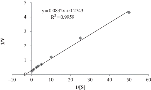
Enzyme kinetic parameters of A. mellea β-glucosidase were obtained by measuring the rate of pNPG hydrolysis at various substrate concentrations ranging from 0.02 to 10 mM in the standard reaction mixture (at 50°C in 50 mM acetate buffer, pH 4.0). The Michaelis–Menten constant (K m) and maximum velocity (V max) values were determined from the Lineweaver–Burk plot using Microsoft Excel software.
Effect of Some Metal Ions on the β-Glucosidase Activity
The effect of metal ions on the enzyme activity was separately investigated by adding chloride salts of Na+, Li+, Mg2+, Mn2+, Zn2+, Co2+, Ca2+, and Cu2+ directly to the standard reaction mixture in a final concentration of 1 mM and 5 mM. Enzyme activity determined in the absence of metal ion was defined as 100%.[ Citation 14 ,Citation 17 ]
Effect of Some Chemicals on the Enzyme Activity
To study the effect of some chemicals on the enzyme activity, phenylmethanesulfonylfluoride (PMSF) and dithiothreitol (DTT) were separately added to the standard reaction mixture in the final concentration of 10 and 25 mM. In addition, the effect of SDS on the enzyme activity was investigated in the final concentration of 0.25 and 0.75%. The percentage residual activities were expressed by comparison with standard assay mixture with no chemical added.[ Citation 14 ,Citation 18 ]
RESULTS AND DISCUSSION
Partial Purification of Armillaria mellea β-Glucosidase
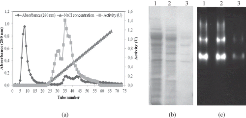
In this study, a β-glucosidase from the fruiting body of A. mellea was partially purified with ion-exchange chromatography (). The enzyme activity and total protein concentration were determined for all fractions collected. The fractions with the highest β-glucosidase activity and the relatively lower protein contents were pooled and concentrated. The β-glucosidase was partially purified 41.1-fold with an overall enzyme yield of 9.1%, and a specific activity of 1.150 U/mg (). It was previously reported that a β-glucosidase from the fruiting body of Lepista flaccida was partially purified 5.0-fold with 30–40% ammonium sulfate saturation.[ Citation 20 ] It was also reported that recombinant Periconia sp. β-glucosidase cloned into Pichia pastoris KM71 was purified 1.1-fold by a single step gel-filtration (Superdex 200).[ Citation 21 ] The purification factor values of β-glucosidases from apple seed, vanilla bean, and soybean have been reported as 47-, 7.2-, and 20-fold, respectively.[ Citation 22 − Citation 24 ]
Table 1 Partial purification of A. mellea β-glucosidase
Figure 1 (a) Purification of A. mellea β-glucosidase by ion-exchange chromatography. (b) SDS-Polyacrylamide gel electrophoresis (PAGE) electrophoresis of the various steps of β-glucosidase isolation (1: crude extract; 2: acetone precipitation; 3: ion-exchange chromatography). (c) Non-denaturing polyacrylamide gel electrophoresis. β-Glucosidase bands were visualized under UV light after necessary procedures (1: crude extract; 2: acetone precipitation; 3: ion-exchange chromatography).
SDS Polyacrylamide Gel Electrophoresis and Activity Staining
The result of SDS polyacrylamide gel electrophoresis is seen in . The number of protein bands was gradually reduced after the acetone precipitation and ion-exchange chromatography, but the number of bands was not one. This shows that the enzyme was purified partially. After the non-denaturing polyacrylamide gel electrophoresis, β-glucosidase bands were visualized under UV light. The presence of more than one band could be attributed to the presence of isoenzymes (). Existing of isoenzymes for β-glucosidases has been previously reported for other organisms such as Lycoperdon pyriforme, L. flaccida, and oat seeds.[ Citation 14 ,Citation 20 ,Citation 25 ]
Determination of Substrate Specificity
β-glucosidase activity was assayed separately by using pNPG, p-nitrophenyl-β-D-mannopyranoside, p-nitrophenyl-β-D-galactopyranoside, and p-nitrophenyl-β-D-cellobioside as substrates. While the highest activity was observed in the presence of pNPG, no activity was found with p-nitrophenyl-β-D-mannopyranoside and p-nitrophenyl-β-D-galactopyranoside (). It was reported that β-glucosidases from Periconia sp. and Termitomyces clypeatus had maximum activity in the presence of pNPG.[ Citation 21 ,Citation 26 ]
Table 2 Substrate specifities of A. mellea β-glucosidase
pH Optimum and Stability
β-Glucosidase activity of the A. mellea increased as a function of pH from 2.0 to 4.0 and decreased gradually after this value (). While the optimum pH was 4.0, more than 80% of maximal activity was also observed between pH 3.0 and 5.0. Similar acidic optimum pH results were also reported for L. pyriforme Aspergillus niger and Metschnikowia pulcherrima. [ Citation 14 ,Citation 27 ,Citation 28 ] The trend for higher specific activity at pH values lower than the optimum of 4.0 reported for most of the β-glucosidases might be due to an acid-base catalytic mechanism.[ Citation 29 ]
Figure 2 (a) pH activity of A. mellea β-glucosidase. (b) pH stability profiles of A. mellea β-glucosidase. (Color figure available online.)
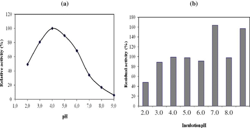
It was determined that A. mellea β-glucosidase was quite stable at pH values between 3.0 and 6.0, and also pH 8.0 after 24 h incubation at 4°C. It retained approximately 90% of its original activity at these values (). Nevertheless, the enzyme was less stable at pH 2.0. Residual β-glucosidase activity increased to 164% and 157% after 24 h incubation at pH 7.0 and 9.0, respectively. Similarly, L. flaccida β-glucosidase retained over 90% of its original activity at pH 4.0 and 5.0 after 24 h incubation at 4°C.[ Citation 20 ] L. pyriforme β-glucosidase was quite stable at pH values between 3.0 and 9.0, and retained over 85% of its original activity at 4°C after 24 h incubation.[ Citation 14 ] Also, β-glucosidase from Periconia sp. retained more than 85% of its maximal activity after incubated at pH 7–10.[ Citation 21 ] It was reported that 90% of A. oryzae β-glucosidase activity remained after 17 h incubation at pH 5.0–7.0 at 30°C. However, only about 70% of the activity remained at pH 4.0.[ Citation 30 ] More than 70% of the original activity of purified Fusarium proliferatum β-glucosidase remained after 24 h incubation at pH 4.0–6.5 at 4°C.[ Citation 31 ]
Temperature Optimum and Thermal Stability
Optimum temperature of A. mellea β-glucosidase was determined as 50°C at pH 4.0 (Fig. 3a). It was reported previously that β-glucosidases from P. pastoris and L. flaccida had optimum temperatures at 40 and 60°C, respectively.[ Citation 16 ,Citation 20 ] It was also reported that L. pyriforme, A. niger, Stachybotrys strain, and Volvariella volvacea β-glucosidases had optimum temperatures of 50°C.[ Citation 14 ,Citation 27 ,Citation 32 ,Citation 33 ]
Figure 3 (a) Temperature-activity of A. mellea β-glucosidase. (b) Thermal-stability profiles of A. mellea β-glucosidase. (Color figure available online.)
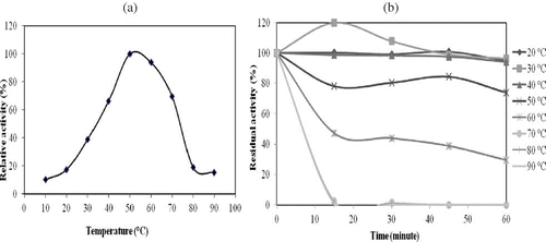
The stability of A. mellea β-glucosidase toward temperature is given in Fig. 3b. While the enzyme retained more than 95% of its original activity at 20 and 40°C after 1 h incubation, its activity increased approximately 20% after a 15-min incubation at 30°C. It had also 75 and 30% activity at 50 and 60°C, respectively, but the activity was severely decreased after 15 minutes at temperatures between 70–90°C. It was reported that Periconia sp. β-glucosidase was found to retain approximately 60% of its maximal activity for at least 2 h at 30–60°C. After incubation at 70°C, it retained more than 50% activity for 2 h.[ Citation 21 ] L. pyriforme β-glucosidase retained its full activity between 20 and 40°C after 1 h, but it lost all of its original activity at 50–80°C.[ Citation 14 ] The residual activity of Debaryomyces pseudopolymorphus β-glucosidase after 3 h incubation at 40°C decreased to approximately 30% of the maximum activity.[ Citation 34 ] It was also reported that β-glucosidase from V. volvacealost 70% of its original activity after only a 5-min incubation at 60°C.[ Citation 35 ] Activity of A. oryzae β-glucosidase significantly decreased at temperatures above 40°C when the enzyme was incubated for 4 h.[ Citation 30 ] When L. flaccida β-glucosidase was incubated at temperatures between 10 and 40°C for 30 min, it retained almost 80% of its original activity, but it lost 35% and 37% of its original activity at 50 and 60°C, respectively.[ Citation 20 ]
Enzyme Kinetics
K m and V max of A. mellea β-glucosidase were calculated from Lineweaver–Burk plot as 0.3 mM and 3.6 U/mg protein, respectively (). K m values of β-glucosidases from other organisms for pNPG range from 0.2 to 21.7 mM.[ Citation 36 ] It was also reported that K m values of L. flaccida, Stachybotrys strain, A. fulica, and Caldicellulosiruptor saccharolyticus β-glucosidases were 1.06, 0.27, 0.224, and 0.67 mM, respectively.[ Citation 20 ,Citation 32 ,Citation 37 ,Citation 38 ] Thus, A. mellea β-glucosidase had a comparable K m value for pNPG. K m is a measure of the affinity of an enzyme for a particular substrate and depends on environmental conditions such as pH, temperature, and ionic strength.[ Citation 39 ] For this reason, the K m values of β-glucosidases from different organisms could be different.
Figure 4 Lineweaver-Burk plot of A. mellea β-glucosidase.
Effect of Some Metal Ions on the β-Glucosidase Activity
The effects of some metal ions on the enzyme activity are seen in . All of the ions except Mn2+ had a negative effect on the β-glucosidase activity. Mn2+ activated the enzyme approximately 28% in the final concentration of 5 mM. It was reported that β-glucosidase activity of Termitomyces clypeatus was inhibited approximately 50, 60, and 55% in the presence of Ca2+ (20 mM), Mg2+ (20 mM), and Cu2+ (2 mM).[ Citation 26 ] A β-glucosidase from Periconia sp. was inhibited 70, 50, 30, and 75% by Cu2+, Zn2+, Mn2+, and Ca2+, respectively.[ Citation 21 ] The metal ions can affect the enzyme by increasing or decreasing the activity. Since metal ions may have different coordination numbers, geometry in their coordination compounds, and potentials as Lewis acids, they may behave differently towards proteins as ligands. These differences may also result in metal binding to different sites, and therefore, change the enzyme structure in different ways and affect the enzyme activity.[ Citation 40 ]
Table 3 Effect of some metal ions on the β-glucosidase activity of A. mellea
Effect of Some Chemicals on the Enzyme Activity
To study the effect of DTT, PMSF, and SDS on the β-glucosidase activity of A. mellea, they were separately added to the standard reaction (). While the enzyme was little affected by DTT, it was inhibited 5 and 39% in the presence of 10 and 25 mM PMSF, respectively. In addition, the enzyme activity was almost fully inhibited by SDS. The inhibition of the enzyme by SDS indicates that the integrity of its three-dimensional structure is critical for its catalytic activity. It was reported that β-glucosidases from Periconia sp. and V. volvacea were also severely inhibited by 1% and 0.1% SDS, respectively.[ Citation 21 ,Citation 33 ] L. pyriforme β-glucosidase was also inhibited 100% in the final concentration of 0.75% SDS.[ Citation 14 ] Activity of T. clypeatus β-glucosidase was inhibited approximately 25% in the presence of 20 mM DTT.[ Citation 26 ]
Table 4 Effect of some chemicals on the β-glucosidase activity of A. mellea
CONCLUSION
In conclusion, a pH and thermostable β-glucosidase was partially purified for the first time from A. mellea, an edible mushroom, with ion-exchange chromatography, and then it was biochemically characterized. The purification fold was 41.1. It was determined that A. mellea β-glucosidase was quite pH and thermal stabile. Especially, thermal stability of A. mellea β-glucosidase is advantageous, since reaction at an elevated temperature provides an opportunity for increased solubility of reactants and products, resulting in higher enzymatic activity, as well as higher reaction velocity stemming from lower viscosity. The risk of contamination is also reduced. Because thermostable cellulytic enzymes also have great potential to be used in industrial processes such as food processing, textiles, and bioconversion, A. mellea β-glucosidase could be useful for these industrial applications.
ACKNOWLEDGEMENTS
The authors wish to thank KTU-BAP (Project number 2009.111.02.7) for financial support and Associate Professor Dr. Dursun Yagiz (Selçuk University, Education Faculty, Konya, Turkey) for identifying the mushroom.
REFERENCES
- Webb , E.C. 1992 . Enzyme Nomenclature 1992. Recommendations of the Nomenclature Committee of the International Union of Biochemistry and Molecular Biology on the Nomenclature and Classification of Enzymes , San Diego , CA : Academic Press .
- Esen , A. and Esen , A. 1993 . “ β-Glucosidases: Overview ” . In β-Glucosidases: Biochemistry and Molecular Biology , 1 – 14 . Washington , DC : American Chemical Society . ACS Symposium Series 533
- Zaldivar , J. , Nielsen , J. and Olsson , L. 2001 . Fuel ethanol production from lignocellulose: A challenge for metabolic engineering and process integration . Applied Microbiology and Biotechnology , 56 : 17 – 34 .
- Krisch , J. , Tako , M. , Papp , T. and Vagvölgyi , C. 2010. . “ Characteristics and potential use of β-glucosidases from Zygomycetes ” . In Current Research, Technology and Education Topics in Applied Microbiology and Microbial Biotechnology , Mendez-Vilas, A. . FORMATEX Research Centre: Badajoz, Spain
- Gueguen , Y. , Chemardin , S.P. , Arnaud , A. and Galzy , P. 1998 . “ Investigation of β-glucosidases potentialities of yeast strains and application to bound aromatic terpenols liberation ” . In New Frontiers in Screening for Microbial Biocatalysts , Edited by: Kieslich , K. , van der Beek , C.P. , de Bont , J.A.M and van den Tweel , W.J.J. 149 – 157 . Elsevier: Amsterdam .
- Coenen , T.M. , Schoenmakers , A.E. and Verhagen , H. 1995 . Safety evaluation of β-glucanase derived from Trichoderma reesei: Summary of toxicological data . Food Chemistry and Toxicology , 33 : 859 – 866 .
- Zhang , Z. , Marquardt , R.R. , Wang , G. , Guenter , W. , Crow , G.H. , Han , Z. and Bedford , M.R. 1996 . A simple model for predicting the response of chicks to dietary enzyme supplementation . Journal of Animal Sciences , 74 : 394 – 402 .
- Jun , S.-Y. , Park , K.-M. , Choi , K.-W. , Kyung Jang , M. , Kang , H.Y. , Lee , S.H. , Park , K.H. and Cha , J. 2008 . Inhibitory effects of arbutin-β-glycosides synthesized from enzymatic transglycosylation for melanogenesis . Biotechnology Letters , 30 : 743 – 748 .
- Yang , J.S. , Chen , Y.W. , Feng , X.Z. , Yu , D.Q. , He , C.H. , Zheng , Q.T. , Yang , J. and Liang , X.T. 1989 . Isolation and structure elucidation of Armillaricin . Planta Medica , 55 : 564 – 565 .
- Elvan , H. , Saglam Ertunga , N. , Yildirim , M. and Colak , A. 2010 . Partial purification and characterization of endoglucanase from an edible mushroom, Lepista flaccida . Food Chemistry , 123 : 291 – 295 .
- Keskin , S. , Saglam Ertunga , N. , Colak , A. , Yildirim Akatin , M. , Özel , A. and Kolcuoglu , Y. 2012 . Characterization of a polyphenol oxidase having monophenolase and diphenolase activities from a wild edible mushroom, Russula delica . Asian Journal of Chemistry , 24 : 1203 – 1208 .
- Lowry , O.H. , Rosebrough , N.J. , Farr , A.L. and Randall , R.J. 1951 . Protein measurement with the folin phenol reagent . The Journal of Biological Chemistry , 19 : 265 – 275 .
- Nagaia , T. , Inoueb , R. , Suzukic , N. and Nagashimaa , T. 2009 . Alpha-Amylase from persimmon honey: Purification and characterization . International Journal of Food Properties , 12 : 512 – 521 .
- Yildirim Akatin . 2013, 16(7): 1565–1577. . “ M. Characterization of a β-glucosidase from an edible mushroom ” . In Lycoperdon pyriforme. International Journal of Food Properties
- Laemmli , U.K. 1974 . Cleavage of structural proteins during the assembly of the head of bacteriophage T4 . Nature , 227 : 680 – 685 .
- Turan , Y. and Zheng , M. 2005 . Purification and characterization of an intracellular β-glucosidase from the methylotrophic yeast Pichia pastoris . Biochemistry (Moskow) , 70 : 1363 – 1368 .
- Yildirim Akatin , M. , Colak , A. and Saglam Ertunga , N. 2013, 37, 177–184. . “ Journal of Food Biochemistry ” . In Characterization of an esterase activity in Lycoperdon pyriforme, an edible mushroom
- Yildirim , M. , Colak , A. , Col , M. and Canakci , S. 2009 . A new recombinant phosphotriesterase homology protein from Geobacillus caldoxylosilyticus TK4: An extremely thermo-pH-stable esterase . Process Biochemistry , 44 : 1366 – 1373 .
- Tuncay , D. and Yagar , H. 2011 . Comparison of polyphenol oxidases prepared from different parts of artichoke (Cynara scolymus l.) . International Journal of Food Properties , 14 : 809 – 821 .
- Elvan , H. , Saglam Ertunga , N. , Colak , A. and Yildirim , M. 2011 . β-Glucosidase from an edible mushroom, Lepista flaccida: Partial purification and characterization . Asian Journal of Chemistry , 23 : 1107 – 1111 .
- Harnpicharnchai , P. , Champreda , V. , Sornlake , W. and Eurwilaichitr , L. 2009 . A thermotolerant β-glucosidase isolated from an endophytic fungi, Periconia sp., with a possible use for biomass conversion to sugars . Protein Expression and Purification , 67 : 61 – 69 .
- Yu , H.-L. , Xu , J.-H. , Lu , W.-Y. and Lin , G.-Q. 2007 . Identification, purification and characterization of β-glucosidase from apple seed as a novel catalyst for synthesis of O-glucosides . Enzyme and Microbial Technology , 40 : 354 – 361 .
- Odoux , E. , Chauwin , A. and Brillouet , J.-M. 2003 . Purification and characterization of vanilla bean (Vanilla planifolia Andrews) β-D-glucosidase . Journal of Agricultural and Food Chemistry , 51 : 3168 – 3173 .
- Hsieh , M.C. and Graham , T.L. 2001 . Partial purification and characterization of a soybean beta-glucosidase with high specific activity towards isoflavone conjugates . Phytochemistry , 58 : 995 – 1005 .
- Kim , Y.-W. and Kim , I.-S. 1998 . Subunit composition and oligomer stability of oat β-glucosidase isozymes . Biochimica et Biophysica Acta – Protein Structure and Molecular Enzymology , 1388 : 457 – 464 .
- Pal , S. , Banik , S.P. , Ghorai , S. , Chowdhury , S. and Khowala , S. 2010 . Purification and characterization of a thermostable intra-cellular β-glucosidase with transglycosylation properties from filamentous fungus Termitomyces clypeatus . Bioresource Technology , 101 : 2412 – 2420 .
- Peshin , A. and Mathur , J.M.S. 1999 . Purification and characterization of β-glucosidase from Aspergillus niger strain 32 . Letters in Applied Microbiology , 28 : 401 – 404 .
- Gonzalez-Pombo , P. , Perez , G. , Carrau , F. , Guisan , J. M. , Batista-Viera , F. and Brena , B.M. 2008 . One-step purification and characterization of an intracellular β-glucosidase from Metschnikowia pulcherrima . Biotechnology Letters , 30 : 1469 – 1475 .
- Legler , G. 1990 . Glycoside hydrolases: Mechanistic information from studies with reversible and irreversible inhibitors . Advances in Carbohydrate Chemistry & Biochemistry , 48 : 319 – 323 .
- Horii , K. , Adachi , T. , Matsuda , T. , Tanaka , T. , Sahara , H. , Shibasaki , S. , Ogino , C. , Hata , Y. , Uedaf , M. and Kondo , A. 2009 . Improvement of isoflavone aglycones production using β-glucosidase secretory produced in recombinant Aspergillus oryzae . Journal of Molecular Catalysis B: Enzymatic , 59 : 297 – 301 .
- Su , J.-H. , Xu , J.-H. , Yu , H.-L. , He , Y.-C. , Lu , W.-Y. and Lin , G.-Q. 2009 . Properties of a novel β-glucosidase from Fusarium proliferatum ECU2042 that converts ginsenoside Rg3 into Rh2 . Journal of Molecular Catalysis B: Enzymatic , 57 : 278 – 283 .
- Amouri , B. and Gargouri , A. 2006 . Characterization of a novel β-glucosidase from a Stachybotrys strain . Biochemical Engineering Journal , 32 : 191 – 197 .
- Li , X. , Pei , J. , Wu , G. and Expression , Shao, W. 2005 . purification and characterization of a recombinant β-glucosidase from Volvariella volvacea . Biotechnology Letters , 27 : 1369 – 1373 .
- Villena , M.A. , Iranzo , J.F.U. , Gundllapallı , S.B. , Otero , R.R.C. and Pérez , A.I.B. 2006 . Characterization of an exocellular β-glucosidase from Debaryomyces pseudopolymorphus . Enzyme and Microbial Technology , 39 : 229 – 234 .
- Cai , Y.J. , Buswell , J.A. and Chang , S.T. 1998 . β-Glucosidase components of the cellulolytic system of the edible straw mushroom, volvariella volvacea . Enzyme and Microbial Technology , 22 : 122 – 129 .
- Joo , A.-R. , Jeya , M. , Lee , K.-M. , Sim , W.-I. , Kim , J.-S. , Kim , I.-W. , Kim , Y.-S. , Oh , D.-K. , Gunasekaran , P. and Lee , J.-K. 2009 . Purification and characterization of a β-1,4-glucosidase from a newly isolated strain of Fomitopsis pinicola . Applied Microbiology and Biotechnology , 83 : 285 – 294 .
- Hu , Y. , Luan , H. , Hao , D. , Xiao , H. , Yang , S. and Yang , L. 2007 . Purification and characterization of a novel ginsenoside-hydrolyzing β-D-glucosidase from the China white jade snail (Achatina fulica) . Enzyme and Microbial Technology , 40 : 1358 – 1366 .
- Hong , M.-R. , Kim , Y.-S. , Park , C.-S. , Lee , J.-K. , Kim , Y.-S. and Oh , D.-K. 2009 . Characterization of a recombinant β-glucosidase from the thermophilic bacterium Caldicellulosiruptor saccharolyticus . Journal of Bioscience and Bioengineering , 108 : 36 – 40 .
- Berg , J.M. , Tymoczko , J.L. and The , Stryer, L. 2002 . “ Michaelis-Menten model accounts for the kinetic properties of many enzymes ” . In Biochemistry , New York : W.H. Freeman .
- Ditusa , C.A. , Christensen , T. , Mccall , K.A. , Fierke , C.A. and Toone , E.J. 2001 . Thermodynamics of metal ion binding.1 . Metal ion binding by wild-type carbonic anhydrase. Biochemistry , 40 : 5338 – 5344 .